
Page 514 - IJB-10-4
P. 514
International Journal of Bioprinting 3D-printed variable stiffness scaffolds
hydrogel contracted from the PCL fibers, thereby leaving Cells mainly clustered around the PCL and grew in
empty spaces for cells to infiltrate. During the culture the direction of the fiber. PCL fibers influenced the cell
period in media, the scaffolds swelled to form a hydrogel orientation, forming an elongated cell morphology due
25
again. In turn, this caused the infiltrated cells to get trapped to its rigid structure. Therefore, matrix formation was
by the PCL fibers, limiting proliferation through the gel found to be secreted along the direction of the PCL fibers.
and compromising cell viability. For example, cell viability Furthermore, it is known that PCL lacks bio-adhesive
in the GelMA scaffolds at D1 was at 43.5%, while the sites. Hence, while PCL guides the formation of tissue, the
viability decreased to 38.6% on D21. This limited internal hydrogel within the construct promotes the production
cell proliferation between D1 and D21 may be due to the of ECM, highlighting that cells are influenced by their
lack of pores, restricting nutrient diffusion through the surrounding environment and the importance of using
swollen hydrogel. multi-material constructs.
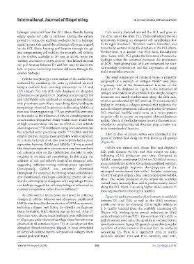
The solid component of meniscal tissue is primarily
Cellular morphology on the surface of the scaffold was 38
evaluated by examining the actin cytoskeletal network composed of a network of collagen fibrils and plays
using a confocal laser scanning microscope on D1 and a primary role in the biomechanical function of the
meniscus. As displayed in Figure 8, the formation of
28
D21 (Figure 7A). On D21, cells displayed an elongated collagen was evident in all scaffolds. Interestingly, collagen
cytoskeleton compared to D1. Scaffolds containing CS/HA formation was mostly predominant along the fiber axis,
and CS/HAMA cells exhibited an elongated morphology which is corroborated by H&E staining. This is an essential
with prominent actin fibers, resembling fibrochondrocyte finding in creating a collagen network that replicates the
morphology observed in previous studies using hMSCs in natural collagen orientation, indicating that the 3D-printed
meniscal tissue engineering. 12,53 Low levels of HA were used PCL meniscal scaffold could serve as a micro-pattern
in this study, as the influence of HA on chondrogenesis is to guide cells to secrete an organized fibrocartilaginous
concentration-dependent. Many studies have found that matrix. This is of particular importance in the meniscus,
at high concentrations HA has no effect or even inhibits whereby its complex internal structure plays a major role
chondrogenesis. 54,55 Nevertheless, using low concentrations in its biomechanical function.
has reported some promising results. 27,56 GelMA and HA After 21 days of culture, GAGs were identified in the
exhibit intrinsic mixing incompatibility when combined. pores of GelMA and along the PCL fibers in all groups
In a study by Levett et al., this immiscibility led to phase (Figure 8).
separation between GelMA and HAMA. It was reported
27
that this phase separation phenomenon may have rendered GelMA was stained with Alcian Blue and displayed
cell adhesion sites on the GelMA less available to cells, fully pink features on D1, and blue streaks on D21,
resulting in rounded cell morphology. In this study, the indicating sGAG production over time. Compared to
addition of HA and HAMA resulted in elongated cells, GelMA, samples containing CS/HA or CS/HAMA stained
suggesting sufficient mixing without phase separation. more positively for sGAGs. CS contains a sulfated polymer,
Consequently, GelMA was uniformly distributed which consequently improves chondrogenesis of the
57
throughout the construct, facilitating robust cell adhesion entrapped mesenchymal stem cells. Samples containing
and proliferation. Hydrogels containing CS/HA are soft, CS at D1 samples display a blue color compared to GelMA
alone. The newly produced sGAG within the scaffolds
and the cells display an elongated cell morphology. Hence, stained more intensely blue and is predominantly found
our findings suggest that cell morphology is influenced by along the PCL fibers, indicating higher sGAG content in
material composition rather than its stiffness. 25 these regions than in the original scaffold.
As cell–material interactions are known to influence Figure 9A and B presents the sGAG content in scaffolds
changes in cellular behavior and phenotype, proliferated between D1 and D21, as well as the GAG secretion
hMCSs were tested for the production of ECM components, profile over time. As observed, CS is highly soluble as
including collagen and GAGs. To identify cartilaginous it is readily leached from the scaffolds containing CS
tissue formation, H&E staining was performed. After 21 (Figure 9C), leading to an overall reduction in GAG
days of in vitro culture, intact hydrogels were still observed content between D1 and D21. The secretion of CS at D1 is
in all groups, and evidence of cartilage tissue formation was high; however, over time, the quantity of secreted sGAG
observed in all samples (Figure 7B). It was observed that steadily decreases. Figure 9D presents more details on the
elongated fibrochondrocytes aligned in rows embedded secretion of sGAG between D14 and D21. In scaffolds
in the newly formed matrix composed of collagen fibers containing CS, there is a significant drop in sGAG
stained pink with H&E. secretion between D14 and D17; however, after D17,
Volume 10 Issue 4 (2024) 506 doi: 10.36922/ijb.3784