
Page 516 - IJB-10-4
P. 516
International Journal of Bioprinting 3D-printed variable stiffness scaffolds
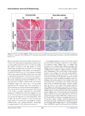
Figure 8. Picrosirius Red staining highlights collagen production (A–F); Alcian Blue staining indicates the production of sulfated glycosaminoglycans
(sGAGs) (G–L). Scale bars: 100 µm. Abbreviations: CS: Chondroitin sulfate; GelMA: Gelatin methacryloyl; HA: Hyaluronic acid; HAMA: Methacrylated
hyaluronic acid.
there is an increase in the secreted sGAG. This increase in Immunohistochemistry between D1 and D21 revealed
GAGs in the media indicates the production and secretion that the different hydrogel compositions could support
of newly synthesized sGAG from the seeded cells within the synthesis of either collagen type I or collagen type
the scaffold. Overall, at D21, the amount of sGAG II (Figure 9E). In hMSC-laden GelMA hydrogels, both
retained and secreted (both normalized to wet weight) collagen types I and II were observed. However, hydrogels
was significantly higher in the constructs containing CS/ containing GAGs displayed a higher intensity of collagen
type I. Furthermore, cells on the surface of all constructs
HA and CS/HAMA (Figure 9A). This finding is consistent produced more collagen than internally in the scaffolds,
with previous reports, whereby GelMA alone may result which is consistent with previous studies. 25,59 This is most
in reduced GAG production. The total sGAG retained in likely due to the lack of cell infiltration and lower nutrient
25
the construct (relative to secreted sGAG) was also higher in concentrations caused by diffusion gradients within the
the samples containing CS/HA and CS/HAMA, i.e., 72%, hydrogel. Cells on the surface of the constructs were able
92%, and 94% for GelMA, GelMA/CS/HA, and GelMA/ to proliferate faster and produce more ECM, as they had
CS/HAMA, respectively (Figure 9B). It was previously better access to nutrients and oxygen. Different collagen
25
reported that HA interacts with ECM components in types are found in each region with varying quantities,
60
matrix retention, particularly when loading is applied. 16,58 with collagen type I predominating within the meniscus.
This may explain the smaller percentage of sGAGs lost Collagen type I is found throughout the meniscus, whereas
to the media in samples containing HA and HAMA collagen type II is exclusively found in the inner two-thirds
compared to GelMA alone. This result suggests that the region. Collagen type I is most abundant in the peripheral
use of GAGs to produce a biomimetic hydrogel provides region of the meniscus and is responsible for 90% of the
composition by dry weight, while other collagens (type II,
cues that may be important for tissue homeostasis. After III, IV, and XVIII) are present in quantities less than 1%.
61
21 days, the net GAG retained in each construct was In the inner region, collagen constitutes approximately
0.02%, 0.21%, and 0.23% of its wet weight for GelMA, 70% of the dry weight, of which 60% is collagen type II and
GelMA/CS/HA, and GelMA/CS/HAMA, respectively, the remaining 40% is collagen type I. Our results indicate
62
i.e., approximately 15–23% of the native meniscus. that a scaffold with regions containing different ECM-like
Volume 10 Issue 4 (2024) 508 doi: 10.36922/ijb.3784