
Page 76 - IJB-10-4
P. 76
International Journal of Bioprinting Unique characteristics of 3D-printed microneedles
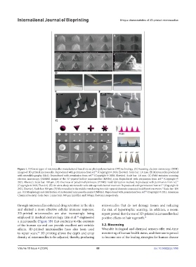
Figure 4. Different types of microneedles manufactured based on vat photopolymerization (VP) technology. (A) Scanning electron microscopy (SEM)
images of 3D-printed microneedle. Reproduced with permission from ref. (Copyright © 2018, Elsevier). Scale bar: 1.0 mm. (B) Microneedles produced
74
with stereolithography (SLA). Reproduced with permission from ref. (Copyright © 2020, Elsevier). Scale bar: 1.0 mm. (C) Field emission scanning
75
electron microscopy (FESEM) images of the 3D-printed hollow microneedles (HMNs) array. Reproduced with permission from ref. (Copyright ©
67
2021, Elsevier). Scale bar: 100 μm. (D) Overview of polydimethylsiloxane (PDMS) mold fabrication method. Reproduced with permission from ref.
71
(Copyright © 2021, Elsevier). (E) An ultra-sharp microneedle with side-opened channel reservoir. Reproduced with permission from ref. (Copyright ©
72
2021, Elsevier). Scale bar: 500 μm. (F) Microneedles in the middle rows having two side-opened channels connected to different reservoirs. Scale bar: 100
73
μm. (G) Morphology and distribution of customized microneedle arrays (CMNAs). Reproduced with permission from ref. (Copyright © 2021, American
60
Chemical Society). Scale bars: 1 mm (top), 300 μm (middle), and 500 μm (bottom), respectively.
through microneedles enhanced drug retention in the skin microneedles that do not damage tissues and reducing
and elicited a more effective cellular immune response. the risk of hypertrophic scarring. In addition, a recent
3D-printed microneedles are also increasingly being report proved that the use of 3D-printed microneedles had
89
employed in medical cosmetology. Lim et al. engineered positive effects on hair regrowth. 91
a microneedle (Figure 5B) that conforms to the contours
of the human eye and can provide excellent anti-wrinkle 3.2. Biosensing
effects. 3D-printed microneedles have also been used Wearable biological and chemical sensors offer real-time
to repair scars. 3D printing allows the depth and array monitoring of human health status, and their use is poised
90
density of microneedles to be adjusted, thereby producing to become one of the leading strategies for human disease
Volume 10 Issue 4 (2024) 68 doi: 10.36922/ijb.1896