
Page 9 - IJB-5-2
P. 9
Zhang, et al.
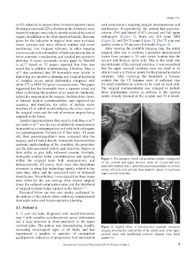
an 8% reduction in surgery time for tumor patients (mean cord compression, requiring surgical decompression and
46 min per case) and 22% reduction in the deformity cases stabilization. Preoperatively, the patient had posterior-
(mean 68 min per case) which directly reduced the cost of anterior (PA) and lateral (LAT) cervical and full spine
surgery in addition to the other reported benefits. Reasons radiographs (Figure 1), brain and full spine MRI
given for the reduction in surgical time were included: (Figure 2), and 3D CT scans (Figure 3). The CT scan was
easier, accurate and more efficient implant and screw used to create a 3D anatomic biomodel (Figure 4).
positioning; less frequent reference to other imaging After viewing the available imaging data, the initial
resources and reduced number of instrumentations due to surgical plan was to perform a posterior instrumented
better anatomic visualization; and detailed pre-operative fusion from occiput to T4 with screw fixation into the
planning. A recent systematic review paper by Martelli occiput and thoracic spine only. Due to the small size
et al. based on 52 papers reported that time was and deformity of the cervical vertebrae, it was considered
[10]
saved due to additive manufacturing. Likewise, Mao et that the upper cervical vertebrae were too small to be
al. also confirmed that 3D biomodels were helpful in able to insert any fixation points for the planned posterior
[8]
improving pre-operative planning and surgical treatment construct. After receiving the biomodel, it became
of complex severe spinal deformities compared with evident that the C2 laminae were of sufficient size
either CT or MRI 3D spinal reconstructions. This paper for small translaminar screws to be used on each side.
suggested that the biomodels were a superior visual aid The surgical instrumentation was changed to include
when confirming the position of an anatomic landmark, these translaminar screws in addition to the fixation
helped the surgeon plan the surgery, facilitated the choice points already planned at the occiput and T3-4 levels.
of internal fixation instrumentation, and improved the
accuracy, and therefore, the safety of pedicle screw
insertion all of which would influence the direct costs of
the surgical cases and the risk of revision surgery being
required in the future.
Another important factor discussed by both Mao et al.
[8]
and Izatt et al. was the use of additively manufactured
[5]
biomodels as a communication tool with both colleagues
and patients/parents. Patients (or if they were <18 years
old, their parents/guardians) were contacted after the
surgery, and all stated that the biomodels improved their
anatomic understanding of the condition; the procedure
and the risks associated with it, and, therefore, improved
their ability to give fully informed consent. Similarly,
biomodels enabled better communication and teaching
within the surgical team both preoperatively and Figure 1. Pre-operative lateral and posterior-anterior radiographs
of the cervical and upper thoracic spine of 12-year-old male
intraoperatively. Of course, there were also limitations (neurofibromatosis type 1, plexiform neuroma posterior to cervical
presented in using this technology mainly related to the spine), which did not provide clear anatomic detail of significant
extra time, labor, and the associated costs of biomodel upper cervical deformity.
manufacture. Nevertheless, it was argued that these issues
were offset by the cost savings from shorter surgical
times, the reduced complication rates, and the likelihood
of surgical revision being required in the future [3,5,7] .
Presented below are two case studies performed by
the authors of this article where additively manufactured
biomodels were used for pre-operative planning.
4.1. Patient A
A 12 year old male, diagnosed with neurofibromatosis
type 1 with complex occipitocervical spinal deformities
and a large neuroma in close proximity to the upper
cervical spine. The patient was demonstrating steadily Figure 2. Sagittal slices of pre-operative magnetic resonance
worsening neurological signs in all limbs and had imaging showing the reduced size of the spinal canal in the upper
experienced a number of episodes of intermittent cervical spine with insufficient posterior element bony detail
quadriparesis indicative of progressive brainstem/spinal (patient A).
International Journal of Bioprinting (2019)–Volume 5, Issue 2 5