
Page 183 - IJB-10-5
P. 183
International Journal of Bioprinting Control nutrients to manipulate fungal growth
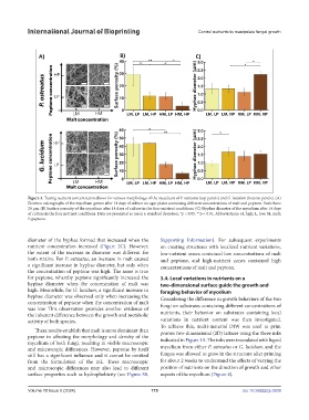
Figure 3. Tuning nutrient concentration allows for various morphology of the mycelium of P. ostreatus (top panels) and G. lucidum (bottom panels). (A)
Electron micrographs of the mycelium grown after 14 days of culture on agar plates containing different concentrations of malt and peptone. Scale bars:
20 μm. (B) Surface porosity of the mycelium after 14 days of culture in the four nutrient conditions. (C) Hyphae diameter of the mycelium after 14 days
of culture in the four nutrient conditions. Data are presented as mean ± standard deviation. *p < 0.05, **p < 0.01. Abbreviations: H, high; L, low; M, malt;
P, peptone.
diameter of the hyphae formed that increased when the Supporting Information). For subsequent experiments
nutrient concentration increased (Figure 2C). However, on creating structures with localized nutrient variations,
the extent of the increase in diameter was different for low-nutrient zones contained low concentrations of malt
both strains. For P. ostreatus, an increase in malt caused and peptone, and high-nutrient zones contained high
a significant increase in hyphae diameter, but only when concentrations of malt and peptone.
the concentration of peptone was high. The same is true
for peptone, whereby peptone significantly increased the 3.4. Local variations in nutrients on a
hyphae diameter when the concentration of malt was two-dimensional surface guide the growth and
high. Meanwhile, for G. lucidum, a significant increase in foraging behavior of mycelium
hyphae diameter was observed only when increasing the Considering the difference in growth behaviors of the two
concentration of peptone when the concentration of malt fungi on substrates containing different concentrations of
was low. This observation provides another evidence of
the inherent difference between the growth and metabolic nutrients, their behavior on substrates containing local
activity of both species. variations in nutrient content was then investigated.
To achieve this, multi-material DIW was used to print
These results establish that malt is more dominant than porous two-dimensional (2D) lattices using the three inks
peptone in affecting the morphology and density of the indicated in Figure 1A. The inks were inoculated with liquid
mycelium of both fungi, resulting in visible macroscopic
and microscopic differences. However, peptone by itself mycelium from either P. ostreatus or G. lucidum, and the
still has a significant influence and it cannot be omitted fungus was allowed to grow in the structure after printing
from the formulation of the ink. These macroscopic for about 2 weeks to understand the effects of varying the
and microscopic differences may also lead to different position of nutrients on the direction of growth and other
surface properties such as hydrophobicity (see Figure S8, aspects of the mycelium (Figure 4).
Volume 10 Issue 5 (2024) 175 doi: 10.36922/ijb.3939