
Page 48 - IJB-10-5
P. 48
International Journal of Bioprinting Bioprinted tumor immune microenvironment
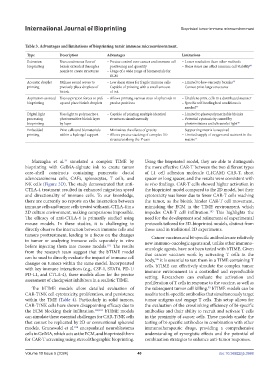
Table 3. Advantages and limitations of bioprinting tumir immune microenvirornment.
Type Description Advantages Limitations
Extrusion Uses continuous flow of – Precise control over cancer and immune cell – Lower resolution than other methods
bioprinting bioink extruded through a positioning and quantity – Shear stress can affect immune cell viability 89
nozzle to create structures – Usage of a wide range of biomaterials for
ECM
Acoustic droplet Utilizes sound waves to – Low shear stress for fragile immune cells – Limited to low-viscosity bioinks 90
printing precisely place droplets of – Capable of printing with a small amount – Cannot print large structures
bioink of ink
Aspiration-assisted Uses aspiration forces to pick – Allows printing various sizes of spheroids in – Unable to print cells in a distributed manner
bioprinting up and place bioink droplets precise positions – Specific self-healing bed conditions is
needed 94
Digital light Uses light to polymerize a – Capable of printing multiple identical – Limited to photopolymerizable bioinks
processing photosensitive bioink layer structures simultaneously – Potential cytotoxicity caused by
bioprinting by layer photoinitiators and ultraviolet light 98
Embedded Print cells and biomaterials – Minimizes the effects of gravity – Supporting matrix is required
printing within a hydrogel support – Allows precise stacking of complex 3D – Limited supply of oxygen and nutrient in the
structures along the Y-axis matrix 77
Mazzaglia et al. simulated a complex TIME by Using the bioprinted model, they are able to distinguish
79
bioprinting with GelMA-alginate ink to create tumor the more effective CAR-T between the two different types
core–shell constructs containing pancreatic ductal of L1 cell adhesion molecule (L1CAM) CAR-T, short
adenocarcinoma cells, CAFs, splenocytes, T cells, and spacer or long spacer, and the results were consistent with
NK cells (Figure 3D). The study demonstrated that anti- in vivo findings. CAR-T cells showed higher activation in
CTLA-4 treatment resulted in enhanced migration speed the bioprinted model compared to the 2D model, but their
and directionality of immune cells. To our knowledge, cytotoxicity was lower due to fewer CAR-T cells reaching
there are currently no reports on the interaction between the tumor, as the bioink hinder CAR-T cell movement,
immune cells and tumor cells treated with anti-CTLA-4 in a mimicking the ECM in the TIME environment, which
2D culture environment, making comparisons impossible. impedes CAR-T cell infiltration. This highlights the
105
The efficacy of anti-CTLA-4 is primarily studied using need for the development and refinement of experimental
mouse models. In these studies, it is challenging to protocols tailored for 3D-bioprinted models, distinct from
directly observe the interaction between immune cells and those used in traditional 2D experiments.
tumors posttreatment, leading to a focus on the changes Cancer vaccines and bi-specific antibodies are relatively
in tumor or analyzing immune cells separately in vitro new immuno-oncologic agents and, unlike other immuno-
before injecting them into mouse models. The results oncologic agents, have not been tested with bTIME. Given
101
from the research team suggest that the bTIME model that cancer vaccines work by activating T cells in the
can be used to directly evaluate the impact of immune cell body, it is essential to test them in a TIME containing T
106
changes on tumors within the same model. Incorporated cells. bTIME can effectively simulate the complex tumor
with key immune interactions (e.g., CSF-1, STAT6, PD-1/ immune environment in a controlled and reproducible
PD-L1, and CTLA-4), these models allow for the precise setting. Researchers can evaluate the activation and
assessment of checkpoint inhibitors in a realistic TIME. proliferation of T cells in response to the vaccine, as well as
The bTIME models allow detailed evaluation of the subsequent tumor cell killing. bTIME models can be
93
CAR-T/NK cell cytotoxicity, proliferation, and persistence used to test bi-specific antibodies that simultaneously target
within the TME (Table 4). Particularly in solid tumors, tumor antigens and engage T cells. This setup allows for
CAR-T/NK cells have shown disappointing efficacy due to the evaluation of the crosslinking efficiency of bi-specific
the ECM blocking their infiltration. 102,103 bTIME models antibodies and their ability to recruit and activate T cells
can simulate these essential challenges for CAR-T/NK cells in the proximity of cancer cells. These models enable the
that cannot be replicated by 2D or conventional spheroid testing of bi-specific antibodies in combination with other
models. Grunewald et al. encapsulated neuroblastoma immunotherapeutic drugs, providing a comprehensive
104
cells in GelMA, which acts as the ECM, and bioprinted them understanding of synergistic effects and the potential of
for CAR-T screening using stereolithographic bioprinting. combination strategies to enhance anti-tumor responses.
Volume 10 Issue 5 (2024) 40 doi: 10.36922/ijb.3988