
Page 45 - IJB-10-5
P. 45
International Journal of Bioprinting Bioprinted tumor immune microenvironment
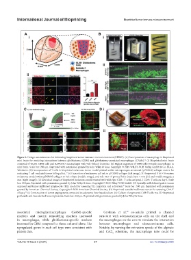
Figure 2. Design considerations for fabricating bioprinted tumor immune microenvironment (bTIME). (A) Incorporation of macrophage in bioprinted
mini brain for modeling interactions between glioblastoma (GBM) and glioblastoma-associated macrophages (GAMs). (i) Bioprinted mini brain
76
consisted of GL261 GBM cells and RAW264.7 macrophages with well-defined locations. (ii) Higher migration of GBM cells towards macrophages in
mini brain. Scale bar: 200 µm. Reprinted with permission granted by John Wiley & Sons. Copyright © 2019 WILEY-VCH Verlag GmbH & Co. KGaA,
Weinheim. (B) Incorporation of T cells in bioprinted melanoma tumor model printed within microporogen-structured (µPOROS) collagen matrix for
evaluating T cell-mediated tumor killing effect. (i) Deposition of melanoma cell ink in µPOROS collagen (left image), 3D-bioprinted B16-F10 murine
77
melanoma model within µPOROS collagen in helix shape (middle image), and side view of printed helix (scale bars: 4 mm [left and middle images]; 2
mm [right image]). (ii) Live/dead images of bioprinted melanoma model treated with wild-type CD8+ T cells and pmel-1 CD8+ T cells on day 3. Scale
bar: 250 µm. Reprinted with permission granted by John Wiley & Sons. Copyright © 2023 Wiley-VCH GmbH. (C) Spatially well-defined gastric tumor
organoid and tumor-infiltrated lymphocyte (TIL) model for assessing TIL migration and activation. Scale bar: 500 µm. Reprinted with permission
80
granted by American Chemical Society. Copyright © 2023 American Chemical Society. (D) Bioprinted vascularized breast cancer for assessing CAR-T
efficacy. (i) Construction of tumor angiogenesis connected to a dynamic flow-based culture. (ii) Culture of engineered CAR-T cells in a 3D-bioprinted
96
perfusable and vascularized tumor spheroids. Scale bar: 200 µm. Reprinted with permission granted by John Wiley & Sons.
associated microglia/macrophages (GAM)-specific Grolman et al. co-axially printed a channel
89
markers and matrix remodeling markers increased structure with adenocarcinoma cells on the shell and
in macrophages, while glioblastoma-specific markers the macrophages on the core to simulate the interaction
increased in GBM compared to those cultured alone. The between macrophages and adenocarcinoma cells.
upregulated genes in each cell type were consistent with Notably, by varying the extrusion speeds of the alginate
patient data. and CaCl solutions, the macrophage tube could be
2
Volume 10 Issue 5 (2024) 37 doi: 10.36922/ijb.3988