
Page 503 - IJB-10-5
P. 503
International Journal of Bioprinting 3D bioprinting of full-thickness skin with a rete ridge structure
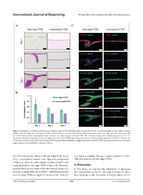
Figure 3. Histological and immunofluorescence staining analyses of full-thickness skin equivalents (FTSEs). (A) Hematoxylin & eosin (H&E) staining
results of the rete ridge and conventional FTSEs. Red dotted boxes indicate well-differentiated keratinocytes at the rete ridge structure. Scale bars: 200
μm. (B) Difference in the interdigitation index between rete ridge and conventional FTSEs. The error bars indicate the standard deviation (n = 4). (C)
Assessment of protein expression in the rete ridge and conventional FTSEs with immunofluorescence staining. High expression of COL17, ITGA6, and
ITGB1 was observed in rete ridge FTSEs (white dotted box and white arrowhead). A higher expression of Kiel 67 (Ki67) was observed at the end of the rete
ridge structure (red dotted box). Scale bars: 100 μm.
K10 was noted in the valleys of the rete ridge FTSE. As for at a depth exceeding 100 μm, a higher number of Ki67
+
Ki67, a discrepancy between rete ridge and conventional cells were observed in rete ridge FTSEs.
FTSEs was observed, with a higher number of Ki67+ cells
being observed in rete ridge FTSEs (Figure 5B). Moreover, 4. Discussion
analysis based on the depth of the skin model (Figure 5C) In this study, we explored the fabrication of advanced
+
revealed a comparable count of Ki67 cells between the flat skin models that mimic the rete ridge structure of native
and rete ridge FTSEs at a depth of 100 μm or less. However, skin, focusing on the discussion and implications of our
Volume 10 Issue 5 (2024) 495 doi: 10.36922/ijb.3961