Page 514 - IJB-10-5
P. 514
International Journal of Bioprinting                          3D model of neurogenesis in Alzheimer’s disease


            porcine skin; and gel strength: ~300 g bloom). GEL (4%   Table 2. Bioprinting parameters.
            w/v)  was  first  dissolved  in  sterile  phosphate-buffered   Parameter           Code
            saline (PBS) on a hot plate magnetic stirrer at 72°C under
            constant agitation of 3 rcf. Once the powder was completely                   1            2
            dissolved, ALG (6% w/v) was added to the solution and   Dimension (mm)
            stirred for 30 min. The final solution was exposed to     X-axis              6            6
            ultraviolet (UV) light for 30 min before printing.    Y-axis                  6            6

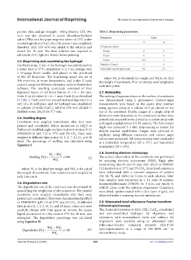
            2.4. Bioprinting and crosslinking the hydrogel        Z-axis                  0.6          1
            For bioprinting, 2 mL of the hydrogel was maintained in   Number of layers    4            6
            a water bath at 37°C, transferred to a 5 mL syringe with   Individual layer height (mm)  0.2  0.2
            a  22-gauge  blunt  needle,  and  placed  in  the printhead
            of the 3D bioprinter. The bioprinting  speed was set at   where Wd  is the initial dry weight and Wd is the final
                                                                                                    f
                                                                          o
            300 mm/min, at room temperature, and under G-code   dry weight of constructs. Four constructs were weighed at
            control, using two different extrusion codes in Pronterface   each time point.
            software. The resulting constructs consisted of four
            deposited layers of cell-laden bioink (6 × 6 × 0.6 mm;   2.7. Wettability
            Code 1) or six layers (6 × 6 × 1 mm; Code 2), as described   The wetting of aqueous drops on the surface of constructs
            in Table 2. Each construct was printed and deposited in a   was characterized using a goniometer. Contact-angle
            well of a 24-well plate, and the hydrogel was crosslinked   measurements were based on the sessile drop method
            in calcium chloride (CaCl ) solution (2% w/v, diluted in   using aqueous drops at a volume of 5 μL placed on the
                                 2
            distilled water, dH O) for 5–7 min.                top of the construct. Briefly, images of a single drop of
                           2
                                                               deionized water deposited on the construct’s surface were
            2.5. Swelling degree                               periodically acquired (every 2 min) by a custom setup with
            Constructs were weighed immediately after they were   a charged-coupled device (CCD) camera. The first contact
            printed and crosslinked, before incubation in dH O or   angle was measured 5 s after drop-casting to ensure the
                                                      2
            Dulbecco’s modified eagle medium/nutrient mixture F-12   droplet reached equilibrium. Images were collected in
            (DMEM/F12) (pH 7.2) at 37°C and 5% CO . They were   triplicate, using different constructs, and contact angle
                                                2
            weighed at different time points (0, 1, 2, 7, 14, 21, and 28   values were measured. All measurements were conducted
            days). The percentage of swelling was calculated using   at a controlled temperature (25 ± 3°C) and humidified
            Equation I:                                        atmosphere (50 ± 10%).

                                W − Wd o                       2.8. Scanning electron microscopy
                                  f
                   Swelling % () =  Wd o  x 100         (I)    The surface observation of the constructs was performed
                                                               by  scanning  electron  microscopy (SEM).  Right after
                                                               crosslinking (day 0) and 14 days after dH O or DMEM/
                                                                                                 2
               where W  is the final wet weight and Wd  is the initial   F12 incubation at 37°C and 5% CO , bioprinted constructs
                                                                                           2
                      f
                                               o
            dry weight of constructs. Four constructs were weighed at   were  dehydrated  with  a  crescent  sequence  of  acetone
            each time point.                                   (30, 50, 70, and 100%) for 5 min in each solution. After
                                                               that, samples were immersed in a 1:1 ratio of acetone:
            2.6. Degradation rate                              hexamethyldisilazane  (HMDS) for 5 min, and finally in
            The degradation rate of the constructs was determined by   HMDS  alone  until  the solution evaporated.  Constructs
            quantifying the weight loss of dry constructs. The printed   were dried, sputter-coated with a thin layer of gold, and
            constructs were weighed immediately after they were   observed under a scanning electron microscope.
            printed and crosslinked. They were then incubated in dH O
                                                        2
            or DMEM/F12 (pH 7.2) at 37°C and 5% CO . At different   2.9. Attenuated total reflectance-Fourier transform
                                               2
            time points (0, 1, 2, 7, 14, 21, and 28 days), constructs were   infrared spectroscopy
            carefully blotted with filter paper to remove the excess   The chemical structures of ALG, GEL, CaCl , crosslinked
                                                                                                   2
            liquid, maintained in a dry oven at 37°C for 20 min, and   and non-crosslinked hydrogel, Aβ oligomers, and
            reweighed. The degradation percentage was calculated   constructs with neurospheres (with and without Aβ
            using Equation II:                                 oligomers)  were analyzed  using  an  attenuated  total
                                                               reflectance-Fourier  transform  infrared  ATR-FTIR
                              Wd −  Wd                                                                   −1
               Degradation % () =  o   f  x 100        (II)    spectrophotometer at a range of 400–4000 cm  in
                                 Wd o                          transmittance mode.

            Volume 10 Issue 5 (2024)                       506                                doi: 10.36922/ijb.3751

