
Page 518 - IJB-10-5
P. 518
International Journal of Bioprinting 3D model of neurogenesis in Alzheimer’s disease
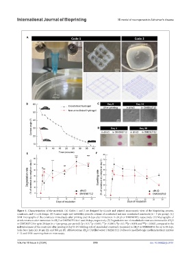
Figure 1. Characterization of the materials. (A) Codes 1 and 2 are designed by G-code and printed: macroscopic view of the bioprinting process,
constructs, and G-code design. (B) Contact angle and wettability periodic scheme of crosslinked and non-crosslinked constructs (n = 3 per group). (C)
SEM micrographs of the constructs immediately after printing and 14 days after immersion in dH O or DMEM/F12, respectively. (D) Macrographs of
2
dried constructs after immersion in dH O or DMEM/F12 for 1 and 28 days, respectively. (E) Degradation rate of crosslinked constructs immersed in dH O
2
2
****
***
##
###
or DMEM/F12 for up to 28 days (n = 4 per group, per period) ( p < 0.5; p < 0.001; p < 0.0001; p < 0.1; p < 0.001; and #### p < 0.0001, compared to the
*
individual mass of the constructs after printing at day 0) (F) Swelling rate of crosslinked constructs immersed in dH O or DMEM/F12 for up to 28 days.
2
Scale bars: 2µm (A); 10 µm (E); and 500 µm (F). Abbreviations: dH O: Distilled water; DMEM/F12: Dulbecco’s modified eagle medium/nutrient mixture
2
F-12; and SEM: scanning electron microscopy.
Volume 10 Issue 5 (2024) 510 doi: 10.36922/ijb.3751