
Page 538 - IJB-10-5
P. 538
International Journal of Bioprinting Biomimetic scaffolds for mandibular repair
enhances the adhesion of mouse pre-osteoblast cells changes are consistent with the findings of Li et al., who
(MC3T3) after being loaded with I-PRF and SDF-1. The reported similar morphological alterations as indicators of
SIT scaffold exhibits excellent mechanical properties early osteogenic differentiation. 33
and biocompatibility and promotes the osteogenic The selection of fields of view in confocal microscopy
differentiation of MC3T3 cells.
can be subjective, and images from a single field of view
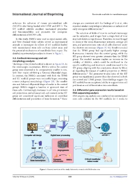
In this study, BMSCs were used as experimental cells, may lack statistical significance. Therefore, we used ImageJ
and New Zealand white rabbits served as experimental to measure the mean fluorescence intensity, average cell
animals to investigate the effects of SIT scaffolds loaded area, and perimeter/area ratio of all cells observed under
with mesenchymal stem cells on bone defect areas and the electron microscope (Figure 2C–E). Results revealed
the potential mechanisms of mandibular bone repair. The that the TPMS group had significantly higher average
detailed experimental procedure is shown in Figure 1. fluorescence intensity than the control group, while the
SIT group showed even greater intensity than the TPMS
3.2. Confocal microscopy and cell group. The marked increase implies an increase in the
morphology analysis viability of BMSCs, which could be attributed to the
The image of the printed scaffold is shown in Figure 2A. In specific scaffolding and treatment conditions used in the
the microscopic examination, BMSCs within the control SIT group, aligning with the conclusions drawn by Shi et
group were observed to be comparatively smaller in size, al. regarding the impact of scaffold properties on BMSC
with their nuclei exhibiting a flattened ellipsoidal shape. differentiation. The perimeter-to-area ratio of the SIT
34
In contrast, the BMSCs associated with both the TPMS group was significantly greater than that observed in both
and SIT scaffold groups were noticeably larger, assuming the control and TPMS groups. These findings suggest the
a more polygonal morphology (Figure 2B). The smaller SIT scaffold has an exceptional ability to support stem cell
size and flattened ellipsoidal shape of nuclei in the control proliferation and differentiation into osteoblasts.
group’s BMSCs suggest a baseline or quiescent state of
these cells. Contrastingly, increases in cell size, prominent 3.3. Differential gene expression results based on
cell projections, and enhanced cell–cell contacts in the SIT RNA sequencing analysis
group are considered significant indicators of osteoblast RNA sequencing analysis was conducted on mesenchymal
differentiation and promotion of bone formation. These stem cells cultured in the SIT scaffolds for 2 weeks to
32
Figure 1. General overview of the study. Schematic created with BioRender.com.
Volume 10 Issue 5 (2024) 530 doi: 10.36922/ijb.4147