
Page 542 - IJB-10-5
P. 542
International Journal of Bioprinting Biomimetic scaffolds for mandibular repair
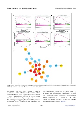
Figure 5. Gene set enrichment analysis (GSEA) and protein-protein interaction network. (A–F) GSEA of differentially expressed genes in SIT scaffolds.
(G) Top 20 protein interaction networks screened by cytoHubba in the Cytoscape.
Osteoblasts in the TPMS and SIT scaffold groups were vascular formation. Compared to the control group, the
mainly concentrated at the interface between the scaffold TPMS and SIT scaffold groups loaded with I-PRF and
and newly formed bone. Additionally, staining of both SDF-1 showed significantly increased new bone tissue and
T and SIT scaffold groups indicated the presence of new
blood vessels (indicated by arrows) distributed within neovascularization within the scaffold structure, with more
the scaffold pores, suggesting that the scaffolds possessed evident bone lacunae, osteoblasts, and vascular networks
appropriate porosity conducive to cell attachment and observed within the scaffolds (Figure 6A).
Volume 10 Issue 5 (2024) 534 doi: 10.36922/ijb.4147