
Page 539 - IJB-10-5
P. 539
International Journal of Bioprinting Biomimetic scaffolds for mandibular repair
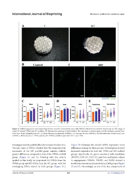
Figure 2. Scaffold appearance and morphology of bone marrow mesenchymal stem cells (BMSCs) observed via confocal microscopy. (A) The image of
actual 3D-printed TPMS and SIT scaffolds. (B) Fluorescence staining of rabbit BMSCs. The cytoplasm is stained green and the nucleus is stained blue.
Scale bars: 30 µm. Magnification: 63×. (C) Mean fluorescence intensity of BMSCs. (D) Average cell area of BMSCs. (E) Perimeter/area (Perim/Area) ratio
of BMSCs. Abbreviations: T: TPMS scaffolds; SIT: TPMS scaffolds loaded with SDF-1 and I-PRF.
investigate how the scaffold affects bone repair mechanisms. Figure 3D illustrates the overall mRNA expression trend
Volcano maps of DEGs revealed that the transcriptomic differences among the three groups. Several genes showed
expression of the SIT scaffold group samples exhibits increased expression in both the TPMS and SIT scaffold
greater differences compared to that of the TPMS scaffold groups. Specifically, the genes associated with osteoblasts
group (Figure 3A and B). Filtering with the criteria (RUNX2, COL1A1, COL1A2, and Osx) and factors related
applied in this study, we pinpointed 413 DEGs from the to angiogenesis (VEGFA, VEGFC, and FGF2) showed a
TPMS group and 853 DEGs from the SIT group, with 316 notable increase in expression in the scaffold groups (Figure
overlapping DEGs shared by both groups (Figure 3C). 3E and F). Interestingly, as one of the key components of
Volume 10 Issue 5 (2024) 531 doi: 10.36922/ijb.4147