Page 544 - IJB-10-5
P. 544
International Journal of Bioprinting                                Biomimetic scaffolds for mandibular repair


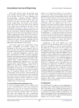
               Alcian blue staining results demonstrated more   weaker or no fluorescence (Figure 7A). As shown in
            active cartilage tissue generation in both the TPMS   Figure 7B, the TPMS scaffold exhibited obvious tissue
            and SIT groups, with the SIT group exhibiting more   ingrowth but few visible microvascular networks, while
            neovascularization,  indicating  potential  additional   the SIT scaffold showed newly formed bone tissue
            advantages of SIT scaffolds in bone repair (Figure 6B).   within the pores and extensive microvascular networks
            Safranin O-fast  green  staining results showed  pink-  (Figure 7C). Within the pores of the SIT scaffold,
            stained collagen fibers and proteins at the junction   particularly under 20× magnification, there  was a
            between the scaffold and newly formed bone compared   stronger c-Jun protein fluorescence signal concentrated
            to the control group, indicating the presence of bone   within the newly formed microvessels, whereas, the
            collagenization (Figure 6C).  Figure 6D illustrates that   overall c-Jun fluorescence intensity in the control and
            the control group exhibited abundant muscle tissue at   TPMS groups was significantly lower. Numerous studies
            the site of bone defect, while the TPMS and SIT groups   have suggested that c-Jun plays a direct or indirect role
            showed no muscle tissue due to the supportive role of the   in angiogenesis. 47,53,54  These clues confirm our previous
            scaffold, suggesting the positive effect of SIT scaffolds in   conjecture that the SIT scaffold promotes the generation
            maintaining the stability of surrounding tissue structure.   of extensive microvascular networks by regulating the
            Mineralized bone tissue was observed in all three groups,   expression of c-Jun protein, and these vascular networks
            represented  by green  coloration, indicating  that the   may facilitate the delivery of oxygen and nutrients,
            bone tissue scaffolds did not significantly inhibit the   promoting the healing and repair of bone defects.
            physiological mineralization of bone repair.          Angiogenesis is the most effective and rapid
               These results suggested that single TPMS scaffolds   method for creating new blood vessels in tissue repair,
            promote osteogenesis and provide support and       like bone regeneration, to provide essential nutrients
            attachment in the defect area, consistent with our   and oxygen for cell function at the site of injury. The
            previous studies.  SIT scaffolds retain the aforementioned   clinical translation of various bone scaffolds made
                         10
            advantages in promoting repair, with an especially better   from different materials has been primarily impeded by
            effect on promoting angiogenesis. Previous studies   inadequate vascularization within the scaffolds and poor
            have demonstrated that SDF-1 possesses angiogenic   osteogenic  activity resulting  from the  differentiation of
            properties, mediating angiogenesis by stimulating human   osteogenesis-related precursor cells, despite extensive in
                                                                                   55–57
            vascular endothelial cells to form microvessels. 49,50  SDF-  vitro and in vivo studies.   We enhanced angiogenesis
            1/CXCR4  exerts  a  regulatory  effect  on  the  JNK/c-Jun   through 3D porous structure design and the delivery of
            pathway, enhancing  the  expression of  c-Jun,  which   angiogenic-related factors. c-Jun plays a significant role
            promotes blood vessel formation. 51,52  These indications   in the AP-1 transcription factor, interacting with and
            suggest that the superior angiogenic effect of SIT is   stimulating the transcription of TRE/AP-1 sequences.
            likely due to SDF-1. SDF-1 promotes angiogenesis by   We found it to be highly expressed in new blood vessels.
                                                               Studies have shown that AP-1 subsequently increases
            upregulating the expression of c-Jun. Dense bone tissue   vascular endothelial growth factor (VEGF), promoting
            and adequate blood  supply are  crucial  for  the effective   endothelial cell proliferation and affecting angiogenesis.
                                                                                                            58
            repair of mandibular defects, and I-PRF containing   In human umbilical vein endothelial cells, c-Jun protein
            various growth factors and SDF-1 with chemotactic and   can induce the expression of downstream genes such as
            recruitment functions play significant roles in stimulating   VEGF.  Since angiogenesis strategies can enhance bone
                                                                    59
            the uniform formation of dense bone tissue and vascular   resorption and formation, 55,60,61  we infer that the superior
            regeneration within the scaffold. Furthermore, no signs   angiogenic effect of SIT scaffolds indirectly facilitate
            of infection or inflammation were observed in any of the   osteogenic repair in the defect area.
            scaffold groups or control groups.
            3.5. Immunofluorescence staining of c-Jun          4. Conclusion
            To further investigate whether the SIT scaffold exerts   Both TPMS and SIT scaffolds promote angiogenesis in
            its role in promoting bone defect repair through the   mandibular defects. TPMS scaffolds loaded with I-PRF and
            regulation of c-Fos/c-Jun, we conducted c-Jun protein-  SDF-1, also known as SIT scaffolds, are more effective at
            specific immunofluorescence staining on mandibular   promoting angiogenesis than pure TPMS scaffolds, solving
            bone tissue from New Zealand rabbits. We observed   the problem of insufficient scaffold vascularization. The
            that under DAPI staining, the nuclei of mandibular   enhanced osteogenic efficiency and angiogenesis observed
            bone tissue cells in all three groups exhibited typical   with the SIT scaffolds may be related to the MAPK pathway
            blue fluorescence, while the cytoplasmic regions showed   and the c-Fos/c-Jun (AP-1) complex.


            Volume 10 Issue 5 (2024)                       536                                doi: 10.36922/ijb.4147

