Page 555 - IJB-10-5
P. 555
International Journal of Bioprinting                                    3D bioprinting of collagen hydrogels


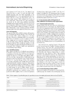
            and incubated at 37 °C with 5% CO . Cell adhesion was   of differentiation-related genes in HFF-1 cells. The 2 −ΔΔCT
                                          2
            observed on days 1, 4, and 7. At each time point, CML-  method, normalized to the housekeeping gene (β-actin)
            scaffold-containing  cells  were fixed  sequentially in  4%   expression levels, was employed to determine the relative
            paraformaldehyde for 10 mins, permeabilized with 0.1%   expression levels of target genes. The primer sequences for
            triton X-100 for 5 mins, and then blocked with a 1%   myofibroblast genes are provided in Table 1.
            bovine serum albumin (BSA) solution for 30 mins at
            room temperature. Subsequently, the actin cytoskeleton   2.7. In situ extrusion with a 3D bioprinter of
            was stained in the dark using phalloidin-tetramethyl   CML-scaffold for full-thickness skin regeneration
            rhodamine isothiocyanate (Solarbio, China) for 60 mins at   2.7.1. Animal model construction
            room temperature. Nuclei were stained by incubating with   Sprague-Dawley (SD) rats were selected for full-thickness
            Hoechst 33258 (Solarbio, China) for 20 mins at 37 °C in the   skin regeneration due to their larger size, which facilitates
            dark. Subsequently, a laser confocal microscope (FV3000;   more extensive surgical procedures and handling of larger
            Olympus, Japan) was used to capture the fluorescence images.  wound areas, allowing for a more accurate assessment of
            2.6.6. Cell migration                              wound healing and biomaterial interactions. Their size also
            The capacity of the CML-scaffold to enhance cell migration   enhances the evaluation of printability and performance of
            was evaluated by quantifying changes in the scratched cell   multi-layer printed scaffolds, enabling better visualization
            area over time. HFF-1 cells at a density of 1 × 10  cells/  and effectiveness in wound repair. Additionally, the use of
                                                     5
            mL were cultured in 6-well plates with 2 mL of medium.   SD rats is well-established in similar studies, providing a
            After 24 hrs of incubation in a CO  incubator (37 °C, 5%   reliable foundation for comparison and validation of the
                                        2
            CO ), a blank area was artificially created at the bottom   findings. This approach has offered valuable insights into
               2
            of the well plate using a white tip. Following a 24-hour   both the wound-healing process and the performance of
            incubation with the CML-scaffold, the migration of cells   the biomaterials.
            in the well plates was observed using inverted fluorescence   Sixty male SD rats, weighing between 150 and 200
            microscopy (IX53; Olympus, Japan) and quantitatively   g, were sourced from the Lanzhou Veterinary Research
            analyzed using ImageJ software.                    Institute (China). They were fed a standard laboratory
                                                               diet. All surgical procedures were performed following the
            2.6.7. Cell differentiation                        guidelines approved by the Ethics Committee of the College
            HFF-1 cells, at a density of 1 × 10  cells/mL, were cultured in   of  Chemistry and  Chemical  Engineering at  Lanzhou
                                     5
            6-well plates containing 2 mL of medium. Following 24 hrs   University (No. G09, 20220711). Prior to the modeling
            of incubation in a CO  incubator (37 °C, 5% CO ), the CML-  procedure, rats underwent anesthesia via intraperitoneal
                                                 2
                            2
            scaffold was placed in 6-well plates with adherent HFF-1   injection of 10% sodium pentobarbital (0.3 mL/100 g).
            cells for seven days. Subsequently, cell differentiation was   Subsequently, the rats’ dorsal fur was shaved, and the area
            assessed by extracting total RNA using an RNA isolation   was cleaned with 0.9% NaCl solution before being sterilized
            kit (AG21101; Accurate Biology, China). RNA samples’   with iodophor. A circular region with a 1.0 cm diameter
            concentration and purity were initially determined with   was  delineated  on  the  back. Using surgical scissors, the
            a spectrophotometer  (Implant,  Germany). Subsequently,   upper skin tissue was entirely excised to create a circular
            cDNA was synthesized using the PrimeScript RT reagent   full-thickness skin defect wound, reaching deep into the
            Kit with gDNA Eraser (Takara, Japan) and analyzed with   fascia. Hemostasis procedures were then carried out.
            TB Green Premix Ex Taq II (Takara, Japan). Reverse-
            Transcription Quantitative Polymerase Chain Reaction   2.7.2. In situ 3D printing of CML-scaffold
            (RT-qPCR) experiments were conducted using a qPCR   The rats were randomly divided into the control and CML-
            system (Mx 3005P; Agilent, USA) to assess the expression   scaffold groups. The wounds in the control group were left


            Table 1. Primer sequence of myofibroblast genes for quantitative reverse transcription polymerase chain reaction (RT-qPCR).
             Gene                                                  Sequence
                                             Forward (5ʹ–3ʹ)                        Reverse (5ʹ–3ʹ)
             α-SMA                      CTGCTGAGCGTGAGATTGTC                   CTCAAGGGAGGATGAGGATG
             Vimentin                   GGCTCGTCACCTTCGTGAAT                  GAGAAATCCTGCTCTCCTCGC
             Col-I                      TTCACCTACAGCACGCTTGT                   TTGGGATGGAGGGAGTTTAC
             Col-III                   GGTCACTTTCACTGGTTGACGA                 TTGAATATCAAACACGCAAGGC


            Volume 10 Issue 5 (2024)                       547                                doi: 10.36922/ijb.4069

