
Page 558 - IJB-10-5
P. 558
International Journal of Bioprinting 3D bioprinting of collagen hydrogels
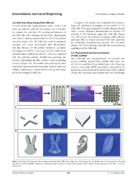
3.2. Real-time 3D printing of the CML-Ink Complex CAD models were employed for extrusion-
A CAD model with a grid structure (8.00 × 8.00 × 2.00 based 3D printing to investigate the printability of the
mm) was selected, with the line distance set at 500 μm, CML-Ink. Printing pentagonal and heart-shaped patterns
to evaluate the real-time 3D printing performance of with a certain thickness demonstrated the efficient 3D
the CML-Ink. After printing the first layer, photographs printing of 3D structures using the CML-Ink (Figure
3C). The precise 3D printing of complex rabbit patterns
were taken to observe and measure the size of the printed and organ-like ear models indicated CML-Ink’s capability
structure (Figure 3A). The CML-Ink could be uniformly for accurately manufacturing intricate 3D structures
extruded and cured immediately after illumination. (Figure 3D). These findings highlight the broad printing
The line distance of the printed structure, calculated capabilities of the CML-Ink.
by ImageJ, was 500.30 ± 10.42 μm, and the width of the
extruded linear scaffold was 262.63 ± 3.50 μm, consistent 3.3. Physicochemical characterization
with the printing settings. Double-layer printing was of CML-scaffold
The CML-Ink was extrusion-based 3D printed into
utilized to investigate the effect of layer count on printing porous scaffolds, named CML-scaffold, with a pore size
accuracy (Figure 3B). The double-layer printing structure of 1.00 mm, and then freeze-dried (Figure 4A). Scanning
maintained micrometer-level precision without structural electron microscopy (SEM) was used to characterize the
collapse, indicating no impact between the printed layers microscopic dimensions of the freeze-dried CML-scaffold
in the 3D printing of CML-Ink. (Figure 4B), revealing a grid pattern with each side length
Figure 3. Real-time 3D printing of the collagen biomaterial ink (CML-Ink). Single-layer 3D printing (A) and double-layer 3D printing (B) of the grid
structure. (C) 3D-printed five-pointed star- and heart-shaped models. (D) 3D-printed complex rabbit- and ear-shaped structures. Scale bars: 1 mm (A);
500 µm (B).
Volume 10 Issue 5 (2024) 550 doi: 10.36922/ijb.4069