
Page 562 - IJB-10-5
P. 562
International Journal of Bioprinting 3D bioprinting of collagen hydrogels
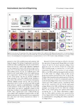
Figure 6. In situ extrusion 3D bioprinting of the collagen biomaterial scaffold (CML-scaffold) for full-thickness skin regeneration. (A) Schematic diagram
of the rat full-thickness skin injury model and treatment method. (B) Gross observation photographs of the wound healing process and the extent of skin
**
repair. (C) Wound healing rate. Data is expressed as mean ± standard deviation (n = 6); p < 0.01. Scale bars: 10mm. Abbreviation: d: Diameter.
appeared in the CML-scaffold group, and epithelial cells Masson’s trichrome staining was utilized to visualize
began to migrate. The epithelial regeneration rates for the the deposition of regenerated collagen fibers in the skin
control and CML-scaffold groups were 25.14 and 48.01%, (Figure 7B and D). On day 4, sporadic collagen fibers
respectively. By day 7, both groups exhibited a significant appeared in the skin wounds of both the control and
amount of granulation tissue, with epithelial regeneration CML-scaffold groups, accounting for 1.15 and 7.39%,
rates of 67.77% for the control group and 83.90% for the respectively. By day 7, the presence of new collagen
CML-scaffold group. On day 14, epithelial tissue had fibers in the CML-scaffold group significantly increased
formed on the wound surface of the CML-scaffold group, to 31.30%, while the collagen fibers in the control
with a regeneration rate of 98.76%, compared to 84.35% group remained less prominent at 4.79%. At day 14, the
in the control group. On day 21, reticular epithelium collagen fiber proportions for each group were 30.84 and
began growing in the CML-scaffold group, reaching a 68.90%, respectively. On day 21, a substantial number of
regeneration rate of 99.45%. Conversely, the control group newly formed collagen fibers were observed in both the
exhibited delayed wound healing, with an epidermal
regeneration rate of 91.64%. By day 28, the CML-scaffold control and CML-scaffold groups, constituting 49.84
group achieved complete re-epithelialization, resembling and 85.71%, respectively. By day 28, the regenerated
normal epidermis. In the control group, the central part collagen fibers in the CML-scaffold group were densely
of the wound was still not completely covered by the arranged, accounting for 95.11%, while the collagen
epidermis, resulting in an epithelial regeneration rate of fibers in the control group were sparsely distributed,
97.79%. These experimental results confirmed that the accounting for 71.15%. These observations suggest
CML-scaffold had a significant reparative effect on full- that the CML-scaffold promotes skin regeneration by
thickness skin injuries, yielding newly formed regenerated enhancing granulation tissue development, accelerating
skin with dense and orderly characteristics similar to re-epithelialization, and guiding the orderly deposition
normal skin. of collagen fibers.
Volume 10 Issue 5 (2024) 554 doi: 10.36922/ijb.4069