
Page 563 - IJB-10-5
P. 563
International Journal of Bioprinting 3D bioprinting of collagen hydrogels
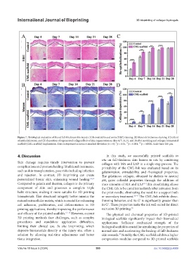
Figure 7. Histological evaluation of the rat full-thickness skin injury: (A) hematoxylin and eosin (H&E) staining, (B) Masson’s trichrome staining, (C) extent
of epithelialization, and (D) deposition of regenerated collagen fibers of skin regeneration on days 4, 7, 14, 21, and 28 after modeling and collagen biomaterial
***
****
scaffold (CML-scaffold) implantation. Data is expressed as mean ± standard deviation (n = 6); p < 0.01, p < 0.001, p < 0.0001. Scale bars: 200 µm.
**
4. Discussion In this study, we successfully printed scaffolds in
situ on full-thickness skin lesions in rats by combining
Skin damage requires timely intervention to prevent collagen with MA and LAP in a single-step process. The
complications and promote healing. Traditional treatments, printability of the CML-Ink was evaluated based on its
such as skin transplantation, pose risks including infection gelatinization, extrudability, and rheological properties.
and rejection. In contrast, 3D bioprinting can create The gelatinous collagen, obtained by dialysis to neutral
personalized bionic skin, enhancing wound healing. 53,54 pH, gains colloidal properties through the addition of
Compared to gelatin and dextran, collagen is the primary trace amounts of MA and LAP. This crosslinking allows
55
component of skin and possesses a complete triple the CML-Ink to be cured immediately after extrusion from
helix structure, making it more suitable for 3D printing the print needle, eliminating the need for a support bath
biomaterials. This structural integrity better mimics the or secondary treatment. 32–41 The CML-Ink exhibits shear-
natural extracellular matrix, which is crucial for enhancing thinning behavior, and its Gʹ is significantly greater than
cell adhesion, proliferation, and differentiation in 3D its G˝. These properties make the ink well-suited for direct
printing applications, thereby improving the performance extrusion 3D printing. 56
and efficacy of the printed scaffolds. 13–16 However, current The physical and chemical properties of 3D-printed
3D printing methods face challenges, such as complex biological scaffolds significantly impact their biomedical
procedures and standalone application difficulties, applications. Sufficient mechanical strength of the
limiting their clinical use. In situ bioprinting, which biological scaffold is crucial for simulating the properties of
deposits biomaterials directly at the injury site, offers a normal skin and accelerating the healing of full-thickness
solution by allowing real-time adjustments and better skin wounds. Notably, the CML-scaffold exhibits a higher
57
tissue integration. compression modulus compared to 3D-printed scaffolds
Volume 10 Issue 5 (2024) 555 doi: 10.36922/ijb.4069