
Page 62 - IJB-10-5
P. 62
International Journal of Bioprinting Medical regenerative in situ bioprinting
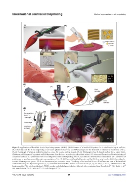
Figure 3. Applications of handheld in situ bioprinting systems (HISBS). (A) Utilization of a handheld bioprinter for in situ bioprinting of scaffolds.
(A, i) Schematic of the in situ bioprinting of cell-laden gelatin methacryloyl (GelMA) hydrogels for the treatment of volumetric muscle loss (VML).
(A, ii) Photograph of a typical scaffold printed on a non-flat porcine skeletal muscle. (A, iii) Photograph of an N-shaped scaffold (three layers thick).
(B) Conceptual diagram of organized scaffold deposition directly into a wound using a handheld printer. (C) Schematic illustration of in vivo printing of
composite scaffolds. (C, i) Schematic view of an integrated camera on the printing pen. (C, ii) Schematic of the material composition. (D) Core/shell-3D
printing via co-axial extrusion. Schematic representation of the (D, i) 3D co-axial handheld printer and the (D, ii) co-axial nozzle. (D, iii) Cartridges for
core/shell-loading in the printer. (E) In situ formation of precursor skin tissue. (E, i) Image of the handheld device. (E, ii) Isometric view of the handheld
instrument during the deposition process. (E, iii) Side view of the handheld device. Scale bars: 5 mm (A, iii); 2.5 cm (E, ii and iii). Abbreviations: HA,
53
54
hydroxyapatite; PCL, poly(caprolactone); VEGF, vascular endothelial growth factor. Adapted with permission from Russell et al. (A), Quint et al.
9
56
(B), Mostafavi et al. (C), Duchi et al. (D), and Cheng et al. (E).
58
Volume 10 Issue 5 (2024) 54 doi: 10.36922/ijb.3366