
Page 23 - IJB-6-1
P. 23
Zhang, et al.
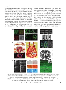
a satisfying artificial lung. The 3D printing has showed two major functions of lung tissues that
helped fabricate lung tissue analog . Lenke et al. are blood flowing and air exchanging. Circulating
[49]
designed a lung model to simulate air-blood barrier moist 10 kPa, 0.5 Hz oxygen gas, the distension of
architecture (Figure 3B). The model consisted the structures, and the shape changes of the concave
of two layers: The endothelial layer containing airway regions could be found. In the meantime,
HUVEC and A549 cells and thin Matrigel layer . they perfuse the deoxygenated red blood cells
[50]
They have not evaluated the functional of the
printed membrane model. However, natural lungs (RBCs) at the blood vessel inlet. Then, they saw
are 3D and can “breathe”. Grigoryan et al. added a the RBCs became clear after passing through the
nontoxic light blocker to improve the z-resolution vessels adjacent to airway. In this work, the high
of DLP 3D printing and fabricated vascularized precision of DLP 3D printing lent them the ability
alveolar model topologies . The alveolar model to fabricate 3D entangled networks in organs.
[9]
A D
B
E
C
Figure 3. Digital light processing 3D printing of artificial tissues: (A) (i) Hepatic structure containing human induced
pluripotent stem cells (hiPSC)-hematopoietic progenitor cells (green) and supporting cells (red) ; (ii) endothelial
[48]
network of prevascularized hepatic tissue , (B) (i) multivascular network; (ii) vascularized lung tissue ,
[47]
[9]
(C) (i) bone tissue engineering scaffold ; (ii) staining of cells encapsulated in bone tissue engineering hydrogel ,
[51]
[52]
(D) hiPSC-derived cardiac tissue on force gauge , (E) spinal cord and its horizontal section .
[53]
[54]
International Journal of Bioprinting (2020)–Volume 6, Issue 1 19