
Page 98 - IJB-6-1
P. 98
Graphene oxide accelerates degradation of poly-l-lactic acid scaffold
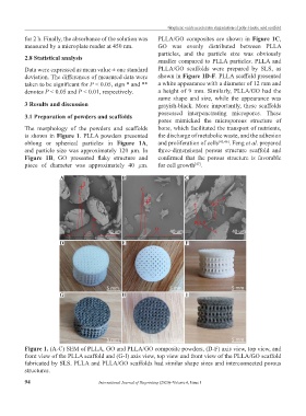
for 2 h. Finally, the absorbance of the solution was PLLA/GO composites are shown in Figure 1C,
measured by a microplate reader at 450 nm. GO was evenly distributed between PLLA
particles, and the particle size was obviously
2.8 Statistical analysis smaller compared to PLLA particles. PLLA and
Data were expressed as mean value ± one standard PLLA/GO scaffolds were prepared by SLS, as
deviation. The differences of measured data were shown in Figure 1D-F. PLLA scaffold presented
taken to be significant for P < 0.05, sign * and ** a white appearance with a diameter of 12 mm and
denotes P < 0.05 and P < 0.01, respectively. a height of 9 mm. Similarly, PLLA/GO had the
same shape and size, while the appearance was
3 Results and discussion grayish-black. More importantly, these scaffolds
possessed interpenetrating micropores. These
3.1 Preparation of powders and scaffolds
pores mimicked the microporous structure of
The morphology of the powders and scaffolds bone, which facilitated the transport of nutrients,
is shown in Figure 1. PLLA powders presented the discharge of metabolic waste, and the adhesion
oblong or spherical particles in Figure 1A, and proliferation of cells [45,46] . Feng et al. prepared
and particle size was approximately 120 μm. In three-dimensional porous structure scaffold and
Figure 1B, GO presented flaky structure and confirmed that the porous structure is favorable
piece of diameter was approximately 40 μm. for cell growth .
[47]
A B C
D E F
G H I
Figure 1. (A-C) SEM of PLLA, GO and PLLA/GO composite powders, (D-F) axis view, top view, and
front view of the PLLA scaffold and (G-I) axis view, top view and front view of the PLLA/GO scaffold
fabricated by SLS. PLLA and PLLA/GO scaffolds had similar shape sizes and interconnected porous
structures.
94 International Journal of Bioprinting (2020)–Volume 6, Issue 1