
Page 89 - IJB-6-2
P. 89
Kolan, et al.
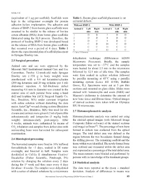
(equivalent of 1 µg per scaffold). Scaffolds were Table 1. Borate glass scaffold placement in rat
kept in the refrigerator overnight for protein calvarial defects.
adhesion before implantation. The adhesion and Without BMP-2 With BMP-2
release of BMP-2 from borate glass scaffolds were Animal # Left Right Animal # Left Right
assumed to be similar to the release of bovine side side side side
serum albumin (BSA) from borate glass scaffolds 1 D D 7 D D
fabricated using the SLS process. Therefore, the 2 D D 8 D D
protocol of loading BMP-2 was developed based 3 D C 9 D C
on the release of BSA from borate glass scaffolds 4 C D 10 C D
that occurred over a period of 4 days. Table 1 5 C C 11 C C
C
C
6
C
C
12
shows the experimental plan of scaffold placement C: Cubic; D: Diamond.
in Sprague Dawley rats.
dehydration technique (EBSciences H2850
2.5 Surgical procedure Microwave Processor). Briefly, the sample
Animal care and use were approved by the temperature was set to ~37°C and the samples
Missouri S&T Institutional Animal Care and Use were heated for about 2.5 min in the microwave
Committee. Twelve 12-week-old male Sprague followed by 12.5 min of idle time. The samples
Dawley rats (~350 g in body weight) were were then soaked in xylene solution followed
anesthetized with a 0.6 ml ketamine/xylazine by paraffin mounting at 45°C using a paraffin
(200 mg ketamine and 20 mg xylazine per 4 ml) mounting system (Leica EG 1150H, Buffalo
abdominal injection. A full-thickness defect Grove, IL). Specimens were cut at 5 µm thin
measuring 4.6 mm in diameter was created in the sections and mounted on glass slides. Slides were
central area of each parietal bone using a hand stained with hematoxylin and eosin (H&E) and
drill and trephine bur (ACE Surgical Supply Co. Masson’s trichrome to determine the amount of
Inc., Brockton, MA) under constant irrigation new bone tissue and fibrous tissue. Optical images
with saline solution without disturbing the dura of stained sections were taken with an Olympus
mater. AutoClip wound closing system (Braintree BX 50 microscope.
®
Scientific, Inc., Braintree, MA) was used for site 2.7 Histomorphometric analysis
closure and animals were given 0.2 ml of penicillin
subcutaneously and ketoprofen (3 mg/kg body Histomorphometric analysis was carried out using
weight) intramuscularly post-surgery. After the stitched optical images (with Microsoft Image
6 weeks, animals were euthanized by means of Composite Editor software) of the stained sections
CO inhalation and samples from defect sites with and ImageJ software . The percentage of new bone
[37]
2
surrounding bone were harvested for subsequent formed in defects was evaluated from the optical
examination. images. The total defect area was defined as the
region between the two boundaries formed during
2.6 Histological processing the drilling process. The remaining scaffold and the
The harvested samples were fixed in 10% buffered tissue within were identified. The newly formed bone
formaldehyde for ~3 days, soaked in DI water was outlined and measured within the defect area
overnight, and bisected in half. Samples were and expressed as a percentage of the total defect area.
decalcified in Cal-Ex II simultaneous Fixative/ The measurements were blinded and performed by
Decalcifier (Fisher Scientific, Pittsburgh, PA) persons with no knowledge of the treatment groups.
solution by changing the solution every 2 days 2.8 Statistical analysis
during the 1 week and then once every 4 days
st
for about ~4 weeks. The samples were dehydrated The data were reported as the mean ± standard
with a series of ethanol solutions by a microwave deviation. Analysis of differences in means was
International Journal of Bioprinting (2020)–Volume 6, Issue 2 85