
Page 73 - IJB-6-4
P. 73
Soman and Vijayavenkataraman
mimic the tumor microenvironment (TME). The of establishing a cancer iPSC depends on the type
commonly used cancer bioprinting methods are: cancer. So far, the successful reprogramming of
Inkject printing, extrusion-based printing, lase- myeloid tumors is established [137-139] . Establishing
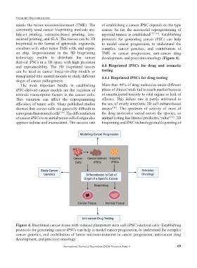
assisted printing, and SLA. The tissues can be 3D protocols for generating cancer iPSCs can help
bioprinted in the format of spheroids, organoids, to model cancer progression, to understand the
coculture with other tumor TME cells, and organ- complex cancer genetics, and contribution of
on chip. Improvements in the 3D bioprinting TME in cancer progression, anti-cancer drug
technology enable to distribute the cancer development, and precision oncology (Figure 4).
derived iPSCs in a 3D space with high precision
and reproducibility. The 3D bioprinted tissues 4.4 Bioprinted iPSCs for drug and cosmetic
can be used as cancer tissue-on-chip models or testing
transplanted into animal models to study different 4.4.1 Bioprinted iPSCs for drug testing
stages of cancer pathogenesis.
The most important hurdle in establishing More than 90% of drug molecules under different
iPSC-derived cancer models are the variation of phase of clinical trials fail to reach market because
intrinsic transcription factors in the cancer cells. of unanticipated toxicity to vital organs or lack of
This variation can affect the reprogramming efficacy. This failure rate is partly attributed to
efficiency of tumor cells. Many published studies the use of overly simplistic 2D cell culture-based
showed that cancer cells are generally difficult to assays [140] . The spectrum of activity of most of
reprogram than normal cells [136] . The differentiation the drug molecules varied across the species, so
of cancer iPSCs to its initial tumor cell of origin also animal testing has limited predictive value [107] . 3D
appears tedious and inconsistent. The success rate bioprinting and iPSC technology enable printing of
Figure 4. Bioprinted cancer tissue with induced pluripotent stem cell (iPSC)-derived cells: Establishing
protocols for generating cancer iPSCs can help to model cancer progression, to understand the complex
cancer genetics, and contribution of tumor microenvironment in cancer progression, anti-cancer drug
development, and precision oncology.
International Journal of Bioprinting (2020)–Volume 6, Issue 4 69