
Page 69 - IJB-6-4
P. 69
Soman and Vijayavenkataraman
commonly used technique in iPSC bioprinting [80,81] 3D bioprinting of peripheral nerve tissue [88,89] for
(Table 2). Extrusion method causes less damage the treatment of peripheral nerve injury.
to the cellular components while printing, as it Human iPSCs are capable of differentiation
uses adjustable mechanical forces with no harsh into many types of specialized cells and have high
treatments for the deposition of the bioink to the value in clinical use. These cells require specific
platform. cell culture media to keep their pluripotent
characteristics intact. The isolation, expansion, and
4.2 Regenerative medicine maintenance of human iPSCs intended for clinical
Autologous iPSCs derived from individuals use should be cultured in xeno-free conditions
provide unlimited source of cells for tissue in compliance with the good manufacturing
regeneration. The unspecialized iPSCs can practice to avoid hypersensitivity reactions after
differentiate and develop to organoids/spheroids transplantation in patients [90,91] . However, many
with specific characteristics of organs in vivo [74,82-84] . conventional protocols of iPSC culture require
These mini-organoids can serve as building blocks to culture in feeder cells. The feeder cells are
for bioprinting of whole organs. Bioengineers usually derived from mouse embryonic fibroblasts
and surgeons are looking for novel methods to (MEFs). The cells are cultured on feeder cells
synthesize artificial skin substitutes that is readily to reduce the genetic instability of the cultured
[89]
available and easily implantable in burn injury cells . Culturing in MEF feeder cells or the
patients [85,86] . Scaffold-free cellular spheroids usage of matrix coating substance (e.g., gelatin
obtained from a coculture of human iPSC-derived or Matrigel) made of animal components make
cardiomyocytes, fibroblasts, and endothelial the iPSCs xeno-positive. Recent introduction of
cells were 3D printed and these cardiac cellular synthetic polymers enables to maintain the iPSC
patches were tested successfully in rat models cultures in xeno-free environment .
[92]
of myocardial infarction . Bioprinted organ Yamanaka factor introduction techniques use
[87]
substitutes such as pancreas, ovary, liver, kidney, different type of retroviral or plasmid vectors
and nervous tissues also will be in high demand in to integrate to the genome of the cell to make it
the near future. Figure 3 shows the workflow of pluripotent. For making clinical grade iPSCs and
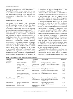
Table 2. Summary of iPSC-based Bioprinting works
Printing Cell source Cells/tissues Bioink used Reference
technique printed
Extrusion iPSCs, BJFF iPSCs Cardiac Collagen I, Matrigel, Gelatin [70]
Human iPSCs Chondrocytes Nano-fibrillated cellulose in alginate [69]
Fibroblasts derived human Germ layers Geltrex [66]
iPSCs
Human iPSCs ((WT I line) Neural construct Matrigel/alginate mixture [68]
Human iPSCs Neural tissues Alginate, carboxymethyl-chitosan, [67]
agarose
Human peripheral blood Pluripotent cells Hydroxypropyl chitin, Matrigel [79]
mononuclear cells derived
iPSCs
Stereolithography Human iPSCs Hepatic Gelatin methacrylate (GelMA), [73]
progenitor cells Glycidal methacrylate-hyaluronic
acid (GMHA)
Laser-assisted Human iPSCs from cord Germ layers Matrigel, Collagen type I, [16]
blood Alginate, Hyaluronic acid
Microvalve-based Human iPSCs Hepatocyte-like Geltrex [19]
cells (HLCs)
International Journal of Bioprinting (2020)–Volume 6, Issue 4 65