
Page 64 - IJB-6-4
P. 64
Applications of 3D bioprinted iPSCs
factors (Oct4, Sox2, and Klf4) , followed by cells. The commonly used bioinks for printing
[7]
epigenetic remodeling of entire genome and two iPSC derived cells are hydrogels derived from
waves of transcriptional events [8,9] . Each cell alginate, carboxymethyl chitosan, agarose, nano-
type in the body require different combinations fibrillated cellulose, hydroxypropyl chitin, gelatin
of transcriptional factors to induce the stemness methacryloyl (GelMA), and Matrigel. Most of
where Oct4 is considered as an indispensable these hydrogels need a crosslinker to give the
core pluripotency gene in the reprogramming final structure of the intended tissue constructs.
process . Exogenous supply of Oct4 alone could Calcium chloride, ultraviolet (UV) radiation,
[10]
convert adult neural stem cells into iPSCs. Recent photo crosslinking, and altered temperatures are
work by An et al. showed that Sox2 and Klf4 used for crosslinking the bioink molecules [25-27] .
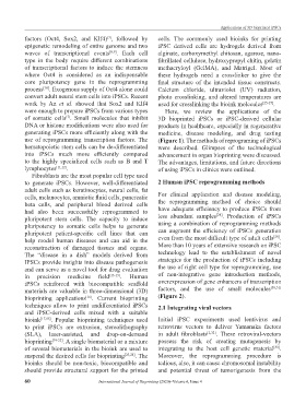
were enough to prepare iPSCs from various types Here, we review the applications of the
of somatic cells . Small molecules that inhibit 3D bioprinted iPSCs or iPSC-derived cellular
[7]
DNA or histone modifications were also used for products in healthcare, especially in regenerative
generating iPSCs more efficiently along with the medicine, disease modeling, and drug testing
use of reprogramming transcription factors. The (Figure 1). The methods of reprograming of iPSCs
hematopoietic stem cells can be de-differentiated were described. Glimpses of the technological
into iPSCs much more efficiently compared advancement in organ bioprinting were discussed.
to the highly specialized cells such as B and T The advantages, limitations, and future directions
lymphocytes [11,12] . of using iPSCs in clinics were outlined.
Fibroblasts are the most popular cell type used
to generate iPSCs. However, well-differentiated 2 Human iPSC reprogramming methods
adult cells such as keratinocytes, neural cells, fat
cells, melanocytes, amniotic fluid cells, pancreatic For clinical application and disease modeling,
beta cells, and peripheral blood derived cells the reprogramming method of choice should
had also been successfully reprogrammed to have adequate efficiency to produce iPSCs from
[28]
pluripotent stem cells. The capacity to induce less abundant samples . Production of iPSCs
pluripotency to somatic cells helps to generate using a combination of reprogramming methods
pluripotent patient-specific cell lines that can can augment the efficiency of iPSCs generation
[29]
help model human diseases and can aid in the even from the most difficult type of adult cells .
reconstruction of damaged tissues and organs. More than 10 years of extensive research on iPSC
The “disease in a dish” models derived from technology lead to the establishment of novel
IPSCs provide insights into disease pathogenesis strategies for the production of iPSCs including
and can serve as a novel tool for drug evaluation the use of right cell type for reprogramming, use
in precision medicine field [13-15] . Human of non-integrative gene introduction methods,
iPSCs reinforced with biocompatible scaffold overexpression of gene enhancers of transcription
materials are valuable in three-dimensional (3D) factors, and the use of small molecules [30,31]
bioprinting applications . Current bioprinting (Figure 2).
[16]
techniques allow to print undifferentiated iPSCs 2.1 Integrating viral vectors
and iPSC-derived cells mixed with a suitable
bioink [17,18] . Popular bioprinting techniques used Initial iPSC experiments used lentivirus and
to print iPSCs are extrusion, stereolithography retrovirus vectors to deliver Yamanaka factors
(SLA), laser-assisted, and drop-on-demand in adult fibroblasts [2,32] . These retroviral-vectors
bioprinting [19-22] . A single biomaterial or a mixture possess the risk of creating mutagenesis by
of several biomaterials in the bioink are used to integrating to the host cell genetic material .
[33]
suspend the desired cells for bioprinting [23,24] . The Moreover, the reprogramming procedure is
bioinks should be non-toxic, biocompatible and tedious, also, it can cause chromosomal instability
should provide structural support for the printed and potential threat of tumorigenesis from the
60 International Journal of Bioprinting (2020)–Volume 6, Issue 4