
Page 67 - IJB-6-4
P. 67
Soman and Vijayavenkataraman
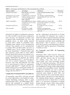
Table 1. Advantages and limitations of the reprogramming methods.
Delivery method Advantages Limitations References
Integrated viral vectors High efficiency, validated in Create insertional mutations, tumor [1,2,31-34]
many cell types induction
Integration free viral vectors. Completely free of vector and Low efficiency of reprogramming, [8,35-38]
For example, Sendai virus, transgene sequences and can reprogramming efficiency varies with
Episomes use in clinical applications, cell types
GMP compatible
mRNAs and miRNAs Faster, high efficiency, absence Multiple transfection required, may [40-44]
of integration, need low input elicit immune response, not evaluated
of starting cells in many cell types
Reprogramming Proteins Foot print-free, cGMP Not evaluated in many cell types, [45-47]
compliant expensive
Small molecules Foot print-free, cGMP Incomplete reprogramming, [48-52]
compliant, economical reprogramming efficiency varies from
cell to cell
attributed to the high-level endogenous expression and the technological advancement in devising
of c-Myc and Klf4 in keratinocytes compared to new generation 3D bioprinters has created a
other cell types. For therapeutic scenarios such whole new field of medical bioprinting that hold
as internal organ reconstruction (e.g., liver, and great promise for artificial tissue/organ printing
peripheral nerve), an ideal starting material for regenerative medicine, disease modeling, and
to create iPSCs should be peripheral tissue. drug testing . Here, in section 4, we describe
[64]
Peripheral tissue samples such as keratinocytes about the commonly used bioprinting techniques
collected from patients can be reprogrammed to print iPSCs and the application of bioprinting
to produce iPSCs and these iPSCs can serve as in health-care field.
a valuable source for the cellular component in
the tissue bioprinting . Peripheral blood cells 4.1 Commonly used iPSC 3D bioprinting
[56]
may represent a good source to derive iPSCs technologies
because taking blood samples from patients are a Bioprinting iPSCs could avoid the ethical and
less invasive clinical procedure. Several research immunological bottle necks of organ printing.
groups standardized the protocols for making iPSCs and iPSC derived cells for developing
iPSC lines from blood cells [57,58] . The iPSC-derived into cartilage, bone, skin, heart, liver, and
cells such as neurons, hepatocytes, osteoblasts, neural tissues have been successfully printed
myocytes, skin cells, and pancreatic beta cells using 3D bioprinting technology. iPSC-derived
are in great demand in clinics for regenerative chondrocytes along with irradiated chondrocytes
medicine applications [59-61] . Small molecules and were bioprinted to cartilage tissues using mixture
growth factors such as recombinant proteins are of alginate and nanocellulose bioink. RegenHu
used in the iPSC cultures to differentiate specific 3D discovery bioprinter was used to print the
lineage cells from iPSCs [62,63] . chondrocytes. Extrusion bioprinting is the most
4 Application of bioprinted iPSCs in healthcare commonly used method to print iPSCs and
organs, followed by SLA, laser-assisted, drop-on
3D bioprinting using iPSCs hold high potential demand, inkject, and microvalve based methods.
for several applications in the health-care sector. The extrusion method applies a pneumatic (air)
Current bioprinting techniques allow to print or mechanical (screw or piston) force to extrude
undifferentiated iPSCs and differentiated iPSC- iPSCs or its derivatives embedded in hydrogel
derived cells embedded in a suitable bioink. bioinks through a nozzle orifice using seamless
Development of novel bioinks, iPSC-derived cells, direct printing . The printing is carried out
[65]
International Journal of Bioprinting (2020)–Volume 6, Issue 4 63