
Page 139 - IJB-10-6
P. 139
International Journal of Bioprinting Fluid mechanics of extrusion bioprinting
Contraction
region
A B C
Figure 8. Visualizations of some unusual behaviors of viscoelastic fluids. (A) Extrudate swell for
Figure 8. Visualizations of some unusual behaviors of viscoelastic fluids. (A) Extrudate swell for viscoelastic fluid issuing from a die (nozzle). Reprinted
100
with permission from ref. Copyright © 2004, John Wiley and Sons. (B) Rod climbing of a viscoelastic fluid over the rotating rod. Adapted from ref. (C)
101
100
viscoelastic fluid issuing from a die (nozzle). Reprinted with permission from ref. Copyright
101
101
Development of secondary flows at contraction flow with low Reynolds numbers (the geometry is similar to that in Figure 3). Reprinted with permission
from ref. Copyright 1994, The Society of Rheology.
102
© 2004, John Wiley and Sons. (B) Rod climbing of a viscoelastic fluid over the rotating rod.
102
Adapted from ref. (C) Development of secondary flows at contraction flow with low Reynolds
thinning and viscoelastic behavior. Additionally, the
biomaterials (listed in Table 4) are natural materials that
numbers (the geometry is similar to that in Figure 3). Reprinted with permission from ref.
103
can have different compositions depending on their source
Copyright 1994, The Society of Rheology. and processing, which affects their rheological behavior.
It is important to note that the absence of thixotropy as
a listed rheological characteristic for a biomaterial in
Table 4 preclude the possibility of thixotropic behavior;
such behavior may emerge at certain concentrations or
compositions. Regarding thixotropy and viscoelastic
behavior, there is a lack of mathematical model fitted
to available test results in the literature, except for an
alginate-based bioink. 112
4. Extrusion multi-material bioprinting
Native tissues possess complex structures composed
of diverse types of materials and cells, with the ECM
organized delicately to fulfill the specific functions of
each tissue and organ. To successfully replicate this
35
complexity in bioprinting, it is necessary to combine
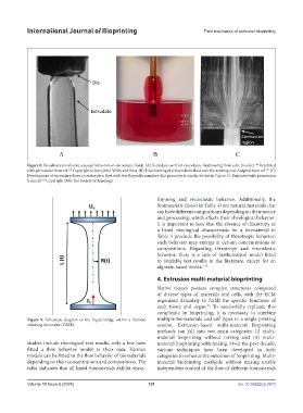
Figure 9. Schematic diagram of the liquid bridge within a filament multiple biomaterials and cell types in a single printing
starching rheometer (FiSER). session. Extrusion-based multi-material bioprinting
methods can fall into two main categories: (i) multi-
material bioprinting without mixing and (ii) multi-
studies include rheological test results, only a few have material bioprinting with mixing. Over the past decade,
fitted a flow behavior model to their data. Various various techniques have been developed in both
Figure 9. Schematic diagram of the liquid bridge within a filament starching rheometer (FiSER).
models can be fitted to the flow behavior of biomaterials categories to enhance the outcomes of bioprinting. Multi-
depending on their concentrations and compositions. The material bioprinting methods without mixing enable
table indicates that all listed biomaterials exhibit shear- independent control of the flow of different biomaterials
Volume 10 Issue 6 (2024) 131 doi: 10.36922/ijb.3973
74