
Page 271 - IJB-10-6
P. 271
International Journal of Bioprinting Skin bioprinting: Keratinocytes and stem cells
conditions, 6 mm diameter pieces were punched out, and with DMEM, supplemented with 10% FCS and 1% P/S,
each scaffold was seeded with 2 × 10 cells dissolved in 100 and added with additional 200 µL reagent suspension. A
5
µL DMEM with 10% FCS and 1% P/S. HaCaTs were left to blank sample without cells was also prepared. The samples
adhere on the scaffolds for 5 min and then transferred to were incubated at 37°C and 5% CO for 4 h, protected from
2
the transwells. light. Approximately 50 µL of reagent solution from both
the lower and upper chambers were merged in a 96-well
After seeding, all transwells were transferred into the
corresponding 24-well plate and covered with 200 µL plate. The absorbance of 100 µL solution was measured
at 450 and 600 nm (background) in a 96-well plate using
DMEM with 10% FCS and 1% P/S (Figure 1). Samples were Multiscan Go and SkanIT RE for Multiscan Go 6.1
incubated at 37°C with 5% CO for 7 days. The medium (Thermo Fisher Scientific, USA) in triplicates.
2
was changed every 48–72 h.
2.3. Material characterization of hydrogels
2.2.3. Cell viability 2.3.1. Printability assay
Live/dead staining of exemplary samples was performed The printability of the different hydrogels (Table 2) was
for demonstration purposes on day 7 for all groups. The evaluated immediately after bioprinting.
transwells were washed in HBSS for 15 min at room
2
temperature. The staining solutions were produced by Scaffolds (1 cm ) were printed using a cylindrical needle
mixing each reagent, 2 µM calcein-acetoxymethyl (calcein- with a 0.25 mm inner diameter at a speed of 5 mm/s. The
AM; Sigma-Aldrich, USA), and 1.5 μM propidium iodide printed scaffolds were placed under a light microscope for
(Sigma-Aldrich, USA), with the cell culture medium. optical magnification. The diagonal length of five crossing
Subsequently, the samples were incubated in calcein-AM points in the printed scaffold was measured (Figure 2).
suspension at 37°C and 5% CO for 30 min, followed by As the inner diameter is 250 µm, the ideal length of the
2
incubation at room temperature in propidium iodide intersection would be 353.55 µm. The diagonal crossing
solution for 5 min. Samples were protected from light and ratio (DCR) of each intersection was determined by
washed twice with HBSS. Microscopic examination in dividing the ideal length by the mean real values of each
100 magnification was conducted using Olympus IX83 crossing point.
(CellSens software; Olympus, Japan).
2.3.2. Cryo-scanning electron microscopy
The WST-8 Assay (PromoCell GmbH, Germany) was The samples were rapidly frozen in slushed nitrogen at
used for measuring the metabolic activity. On days 1, 4, −210°C after placing them between aluminum plates
and 7, three replicates of each group were examined. The (diameter: 3 mm) with a 2 mm notch for sample fixation.
reagent solution was prepared by mixing CCVK1 reagent Subsequent transfer steps were performed at −140°C with
with DMEM, supplemented with 10% FCS and 1% P/S, in a EM VCT100 cryo-shuttle (Leica Microsystems, Germany).
ratio of 1:11. The transwells were transferred into a new 24- To generate a freshly fractured hydrogel surface, one of the
well plate, containing 500 µL CCVK1 reagent suspension aluminum plates was knocked off and freeze-etched for 15
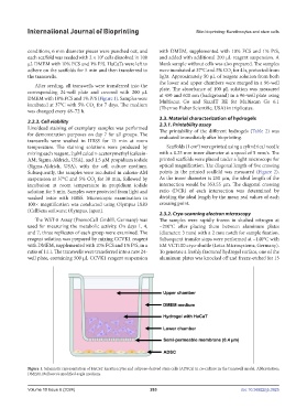
Figure 1. Schematic representation of HaCaT keratinocytes and adipose-derived stem cells (ADSCs) in co-culture in the transwell model. Abbreviation:
DMEM, Dulbecco’s modified eagle medium.
Volume 10 Issue 6 (2024) 263 doi: 10.36922/ijb.3925