
Page 274 - IJB-10-6
P. 274
International Journal of Bioprinting Skin bioprinting: Keratinocytes and stem cells
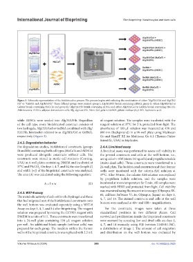
Figure 3. Schematic representation of the biofabrication process. The process begins with selecting the combination of either “Alg/HA/Gel and Alg/HA/
Fib” or “GelMA and Alg/HA/Fib.” Three different groups were printed: group I: Alg/HA/Fib bioink containing ADSCs; group II: either Alg/HA/Gel or
GelMA bioink containing HaCaTs; and group III: Alg/HA/Fib bioink containing ADSCs and either Alg/HA/Gel or GelMA bioink containing HaCaTs.
Abbreviations: ADSCs, adipose-derived stem cells; Alg, alginate; Fib, fibrin; Gel, gelatin; GelMA, gelatin methacryloyl; HA, hyaluronic acid.
while ADSCs were seeded into Alg/HA/Fib. Regardless of reagent solution. The samples were incubated with the
of the cell type, every biofabricated construct consists of reagent solution at 37°C for 2 h, protected from light. The
two hydrogels, Alg/HA/Gel or GelMA combined with Alg/ absorbance of 100 µL solution was measured at 450 and
HA/Fib, hereinafter referred to as Alg/HA/Gel or GelMA, 600 nm (background) in a 96-well plate using Multiscan
respectively (Figure 3). Go and SkanIT RE for Multiscan Go 6.1 (Thermo Fisher
Scientific, USA) in triplicates.
2.4.2. Degradation behavior
For degradation studies, biofabricated constructs (groups 2.4.4. Live/dead assay
Ib and IIb) containing both cell types (HaCaTs and ADSCs) A live/dead assay was performed to assess cell viability in
were produced alongside constructs without cells. The the printed constructs and cells at the well bottom, i.e.,
constructs were stored in sterile cell strainers (Corning, using calcein-AM (stains living cells) and propidium iodide
USA) in 6-well plates containing DMEM and incubated at (stains dead cells). Three constructs were transferred to a
37°C and 5% CO . On days 1, 4, 7, and 14, the size (length [l] 24-well plate. The biofabricated constructs and their former
2
and width [w]) of the bioprinted constructs was analyzed. wells were incubated with the calcein-AM solution at
The area (A) was calculated using the following equation: 37°C. After 30 min, the calcein-AM solution was replaced
by propidium iodide solution, and the samples were
A = l × w (II) incubated at room temperature for 5 min. All samples were
washed with HBSS and protected from light. Cell viability
was examined using fluorescent microscopy (Olympus IX-
2.4.3. WST-8 assay 83, cellSens Software V1.16; Olympus, Japan) on days 1,
The metabolic activity of cells within the hydrogel and those 4, 7, and 14. The stained constructs and cells at the well
that had migrated out of the biofabricated constructs onto bottom were analyzed at 40 and 100 magnifications.
the well bottom was evaluated separately using a WST-8
Assay on days 1, 4, 7, and 14 after bioprinting. The reagent For the constructs, images were taken at three
solution was prepared by mixing the CCVK1 reagent with standardized positions in two different planes. Cell
DMEM in a ratio of 1:11. Three constructs were transferred survival and proliferation inside the bioprinted constructs
into a 24-well plate containing 550 µL reagent solution were assessed by counting live and dead cells on days 1,
per well. An additional blank sample (without cells) was 4, 7, and 14 manually using FIJI (version 2.140.0/1.54f),
prepared for each group. The medium within the former a distribution of Image J. The amount of cell migration
wells of the bioprinted constructs was replaced with 2.2 mL and distribution on the well bottom was evaluated by
Volume 10 Issue 6 (2024) 266 doi: 10.36922/ijb.3925