
Page 278 - IJB-10-6
P. 278
International Journal of Bioprinting Skin bioprinting: Keratinocytes and stem cells
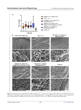
Figure 6. Scanning electron microscopy (SEM) of the different hydrogels (n = 1). (A) Pore sizes measured from the SEM images and displayed as box plots
with whiskers. (B) Exemplary images of the hydrogels in two different magnifications. The magnification of the upper pictures is 1000 (top image) and
5000 (bottom image), the magnification of the lower picture is 5000-fold. Scale bars: 5 µm. Abbreviation: GelMA, gelatin methacryloyl.
Volume 10 Issue 6 (2024) 270 doi: 10.36922/ijb.3925