
Page 281 - IJB-10-6
P. 281
International Journal of Bioprinting Skin bioprinting: Keratinocytes and stem cells
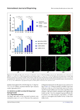
Figure 11. Cell viability of cells at the well bottom. (A) Metabolic activity of HaCaT and adipose-derived stem cells (ADSCs) at the well bottom on
days (d) 1, 4, 7, and 14 (n = 3). (B) Living cells at the well bottom on days 1, 4, 7, and 14 (n = 3). (C–D) Cells at the well bottom from cell-laden Alg/
HA/Gel constructs day 14: HaCaT (top left); ADSCs (bottom right). (E) Cells at the well from cell-laden Alg/HA/Gel constructs on days 1, 4, 7, and 14;
magnification: 40. Scale bars: 200 μm (C); 5 mm (D); and 2 mm (E). Green: living cells; red: dead cells. Data are represented as mean ± standard deviation
(n = 3); *p ≤ 0.05. Abbreviations: Alg, alginate; Gel, gelatin; GelMA, gelatin methacryloyl; HA, hyaluronic acid.
medium. Hydrogels II (Fib), III (Fib/Alg/HA), V (Alg/Gel/ of all groups decreased over the experimental period of 14
HA), and VI (GelMA) reported relatively high measured days (Figure 9A). Statistical analysis revealed a significant
relative light units (RLU). difference in the size of the bioprinted constructs between
Alg/HA/Gel and GelMA at all time points, regardless of cell
3.3. HaCaTs in an ADSC-enriched 3D-bioprinted presence. Cell-laden GelMA constructs were significantly
hydrogel model smaller on day 7 than the comparative group without cells.
3.3.1. Degradation behavior In addition, for both hydrogel compositions, cell-laden
The length and width of the bioprinted constructs were constructs were significantly smaller on day 14 than those
evaluated on days 1, 4, 7, and 14. The size of the constructs without cells.
Volume 10 Issue 6 (2024) 273 doi: 10.36922/ijb.3925