
Page 296 - IJB-10-6
P. 296
International Journal of Bioprinting 3D-Printed Zn/MgHA-PCL for angio/osteogenesis
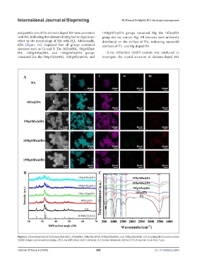
and particle size of the element-doped HA were consistent 15Mg10Zn@HA groups contained Mg; the 10Zn@HA
with HA, indicating that element doping had no significant group did not contain Mg. All elements were uniformly
effect on the morphology of HA with H L. Additionally, distributed on the surface of HA, indicating successful
6
EDS (Figure 2A) displayed that all groups contained synthesis of Zn- and Mg-doped HA.
elements such as Ca and P. The 10Zn@HA, 5Mg10Zn@
HA, 10Mg10Zn@HA, and 15Mg10Zn@HA groups X-ray diffraction (XRD) analysis was conducted to
contained Zn; the 5Mg10Zn@HA, 10Mg10Zn@HA, and investigate the crystal structure of element-doped HA
Figure 2. Characterization of hydroxyapatite (HA), 10Zn@HA, 5Mg10Zn@HA, 10Mg10Zn@HA, and 15Mg10Zn@HA. (A) Scanning electron microscope
(SEM) images and element mapping. (B) X-ray diffraction (XRD) patterns. (C) Fourier transform infrared (FTIR) spectra. Scale bars: 5 μm.
Volume 10 Issue 6 (2024) 288 doi: 10.36922/ijb.4243