
Page 301 - IJB-10-6
P. 301
International Journal of Bioprinting 3D-Printed Zn/MgHA-PCL for angio/osteogenesis
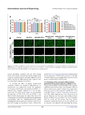
Figure 5. Cell viability and proliferation activity of rat bone marrow mesenchymal stem cells (RBMSCs) in each group of scaffolds. (A) Proliferative activity
of RBMSCs after 1, 3, and 5 days of culture (*p < 0.05, **p < 0.01, ***p < 0.001; n = 4). (B) Live/dead staining images of each group of scaffolds. Scale bars:
500 μm. Abbreviations: HA, hydroxyapatite; ns: non-significant; PCL, polycaprolactone;.
mineral deposition, consistent with the ARS staining results (Figure S4, Supporting Information) demonstrated
results. These results indicated that Mg/Zn double-doped that the expression of OPN, OCN, and COL1 in the Mg/
composite scaffolds promote osteogenic differentiation of Zn double-doped group was higher than that in the control
BMSCs and that the differentiation effect improves with group, consistent with the qPCR results.
increasing Mg ion doping concentration.
3.5. Evaluation of the osteogenic differentiation
To elucidate the effect of Mg/Zn double-doped potential of 3D-printed composite scaffolds in vivo
composite scaffolds on osteogenic differentiation, we Using a rat femoral defect model, the ability of the composite
quantitatively and qualitatively studied the expression scaffolds to promote bone repair was evaluated. H&E and
of osteogenic-related genes and proteins in BMSCs co- Masson staining were used to evaluate bone regeneration
cultured with composite scaffolds for 7 and 14 days, following treatment with composite scaffolds after 1 and
respectively. The qPCR results (Figure 6E) indicated that 3 months (Figures 7 and S6, Supporting Information).
the expression of OPN, OCN, ALP, and RUNX2 in the The H&E results displayed new bone formation in all
Mg/Zn double-doped group was higher than that in the groups. However, the Zn-doped and Zn/Mg-co-doped
control group and positively correlated with the Mg doping groups exhibited more prominent new bone formation
concentration, with the 15Mg10Zn@HA-PCLs group than the control and HA groups. New bone formed
exhibiting the highest expression, consistent with the ALP around the scaffold (the blank area was the scaffold), and
and ARS staining and quantitative results. Western blot its formation was enhanced with increasing Mg doping.
Volume 10 Issue 6 (2024) 293 doi: 10.36922/ijb.4243