
Page 303 - IJB-10-6
P. 303
International Journal of Bioprinting 3D-Printed Zn/MgHA-PCL for angio/osteogenesis
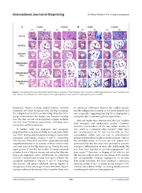
Figure 7. (A) Hematoxylin and eosin (H&E) and (B) Masson staining of bone formation after composite scaffold implantation for 1 and 3 months. Scale
bars: 1000 μm (2×); 400 μm (5×). Abbreviations: HA, hydroxyapatite; N, new bone; PCL, polycaprolactone; S, scaffold.
Meanwhile, Masson staining analysis further validated no significant differences between the scaffold groups,
enhanced new bone formation with Zn/Mg-co-doping. and the collagen type I content in the newly formed bone
The 10Mg10Zn@HA-PCLs and the 15Mg10Zn@HA-PCLs was comparable, suggesting that Mg/Zn-co-doping has a
groups demonstrated the deepest and broadest staining synergistic effect in promoting bone regeneration.
area (the blue and red colors represent collagen synthesis Relevant studies have demonstrated that Zn exhibits
2+
and new bone formation, respectively), indicating more both osteogenic and antibacterial activity. However,
43
calcification and bone maturity.
excessive concentrations of Zn may induce toxicity in
44
To further verify the angiogenic and osteogenic vivo, which is a potential safety hazard. High doses
properties of the composite scaffolds, we conducted α-SMA and concentrations of Zn ions are harmful, as their
and CD31 immunohistochemical staining to assess their accumulation within cells can lead to mitochondrial
angiogenic performance (Figures 8 and S7, Supporting damage and disruption of cellular Zn homeostasis. Hence,
45
Information). The α-SMA and CD31 results demonstrated the controlled release of Zn ions not only reduces their
a significant increase in the number of blood vessels in the cytotoxicity but also harnesses their potential to promote
new bone areas of the Mg-doped group. During the early osteogenic differentiation of stem cells. Additionally, Mg
repair process (1 month), the number of vessels increased ions have been reported to promote angiogenesis and
46
at higher levels of Mg ion doping, indicating that Mg ions bone repair. Therefore, co-doping of HA with Zn and
1,47
have a pro-angiogenic effect. Additionally, we performed Mg not only achieves slow and safe release of Zn ions but
COL1A1 immunohistochemical staining to evaluate the also synergistically enhances angiogenesis to facilitate
osteogenic performance (Figures 8 and S7, Supporting bone repair. Therefore, this study employed a continuous
Information). All groups exhibited varying degrees of sustained release of Zn to ensure its safe release in vivo.
2+
osteogenesis. In the early stages of bone formation, Mg ion Likewise, the addition of Mg can synergistically promote
2+
doping accelerated the process. After 3 months, there were vascular growth and new bone generation.
Volume 10 Issue 6 (2024) 295 doi: 10.36922/ijb.4243