Page 329 - IJB-10-6
P. 329
International Journal of Bioprinting                            3D-printed PPDO/GO stents for CHD treatment


            2.6. Surface morphology analysis of PPDO/GO films   supplemented with 1% penicillin-streptomycin (HyClone,
            and sliding-lock stents                            USA) and 10% fetal bovine serum (Gibco, USA), before
                                                               incubation in a humidified incubator with 5% CO  at 37°C.
               The surface morphology of PPDO/GO films and                                            2
            sliding-lock  stents  was  analyzed  by SEM  using  a  RISE-  The culture medium was refreshed every two days. PPDO/
                                                               GO stents were cut into small pieces (1 × 1 cm), washed
            MAGNA system (TESCAN, Czech Republic) at an        with 75% ethanol solution and deionized water, and
            accelerating voltage of 5 kV. The surfaces were sputtered   sterilized by ultraviolet radiation. After sterilization, the
            with gold before observation.
                                                               samples were placed into 12-well culture plates, immersed
            2.7. In vitro evaluation of hemocompatibility      in DMEM, and incubated for 2 h.
            2.7.1. Hemolysis test                              2.8.2. Cell adhesion and proliferation
            Fresh Sprague–Dawley (SD) rats blood was collected   The HUVECs were seeded on the samples in 12-well
                                                                                          4
            in  heparinized test  tubes.  Red blood cells (RBCs) were   culture plates at a density of 2 × 10  cells/mL and incubated
            obtained by centrifuging the anticoagulated blood at 4000   in the culture medium. After incubation for 1, 3, 5, and
            r/min for 5 min and then removing the supernatant plasma.   7 days, cell viability and proliferation were evaluated by
            The 2% RBC solution was prepared by dispersing the RBCs   a Cell Counting Kit-8 (CCK-8) assay according to the
            in normal saline. PPDO/GO stents were cut into small   manufacturer’s protocol. The culture medium was removed,
            pieces and incubated with 2 mL of the 2% RBC solution   and the samples were washed with PBS. The samples were
            at 37°C for 2 h. After the removal of stents, the suspension   then transferred to a 12-well plate, and CCK-8 reagent
            was centrifuged at 4000 r/min for 5 min. For positive and   (Beyotime, China) was added to each well and incubated
            negative controls, the 2% RBC solution was incubated   for 2 h at 37°C. The absorbance of the supernatant was
            with deionized water and normal saline, respectively.   measured using a microplate reader at 450 nm.
            The absorbance of the supernatant was measured at 545   2.8.3. Cell morphology
            nm  using  a  microplate  reader. The  hemolysis  rate  was   The HUVECs were seeded on the samples in 12-well culture
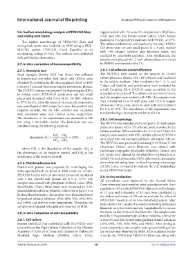
            calculated using the following equation:           plates at a density of 2 × 10  cells/mL and incubated in the
                                                                                    4
                                                               culture medium. After incubation for 1, 3, 5, and 7 days, the
                                  OD − OD                      samples were washed with PBS, and the adhered HUVECs
                   Hemolysis % () =  s    n  ×100 %    (III)
                                  OD − OD                      were fixed with 4% paraformaldehyde (Beyotime, China).
                                     p
                                          n
                                                               The HUVECs were permeabilized using 0.1% Triton X-100
                                                               (Beyotime, China). Actin filaments were stained with
               where  OD   is  the  absorbance  of  the  sample,  OD   is
                       s
                                                       n
            the absorbance of the negative control, and  OD  is the   rhodamine-conjugated phalloidin (Solarbio, China), and
                                                               cell nuclei were stained by 4,6-diamidino-2-phenylindole
                                                    p
            absorbance of the positive control.
                                                               (DAPI; Vector Laboratories, USA). Afterwards, the samples
            2.7.2. Platelet adhesion test                      were observed using laser confocal scanning microscopy
            Platelet-rich  plasma was  prepared  by centrifuging  the   (LCSM; Leica, Germany) to evaluate the cell morphology
            anticoagulated fresh rat blood at 1000 r/min for 10 min.   on the PPDO/GO stents.
            PPDO/GO stents were cut into small pieces and incubated   2.9. In vivo evaluation
            with 1 mL platelet-rich plasma for 2 h at 37°C. The   All procedures were approved by the Animal Ethics
            samples were rinsed with phosphate-buffered saline (PBS;   Committee and performed in strict accordance with their
            BasalMedia, China) three times and immersed in 2.5%   regulations. 3D-printed PPDO/GO filaments with a length
            glutaraldehyde solution (Solarbio, China) for at least 6 h to   of 15 mm and a diameter of 0.1 mm were implanted in
            fix the adhered platelets. The samples were then dehydrated   the abdominal aortas of SD rats to evaluate the effect of
            by gradient ethanol solutions (50%, 60%, 70%, 80%, 90%,   PPDO/GO materials on in vivo endothelialization. After
            and 100%) and dried at room temperature. Thereafter, the   implantation for 2 weeks, the vessels containing embedded
            samples were sprayed with gold and observed by SEM.  filaments were harvested and cut longitudinally to expose

            2.8. In vitro evaluation of cell compatibility     the tissue on the surface of the filaments. The samples were
                                                               fixed by 2.5% glutaraldehyde solution (Solarbio, China) for
            2.8.1. Cell culture                                at least 6 h and dehydrated using gradient ethanol solutions
            Human umbilical vein endothelial cells (HUVECs) were   (50%, 60%, 70%, 80%, 90%, and 100%). After drying at
            sourced from the Type Culture Collection of the Chinese   room temperature, the samples were sprayed with gold on
            Academy of Sciences (China) and cultured in Dulbecco’s   the surface and observed by SEM. After implantation for
            Modified Eagle Medium (DMEM; Gibco, USA),          4 weeks, the PPDO and PPDO/GO filaments, along with

            Volume 10 Issue 6 (2024)                       321                                doi: 10.36922/ijb.4530

