Page 371 - IJB-10-6
P. 371
International Journal of Bioprinting                                           3D-printed NAFLD model


            2.7. CDFDA staining                                of glyceraldehyde-3-phosphate dehydrogenase (GAPDH)
            Bile  canaliculi formation  was evaluated  via the use  of   via the 2 −ΔΔCt  method.
            5(6)-carboxy-2’,7’-dichlorofluorescein diacetate.  The
                                                    27
            CDFDA stock solution was diluted with PBS to create a   2.10. Induction of NAFLD
            5 μM working solution for staining. The samples were   To  induce  NAFLD,  we  used  a  commonly  employed
            washed with PBS before being exposed to the CDFDA   induction method and fatty acid ratios, creating a
            working solution at 37°C for 20–30 min. Subsequently, 10   lipid culture medium composed of a base medium
            μg/mL DAPI was added, and the samples were incubated   supplemented with 5.5 mM glucose and a mixture of free
                                                                                               27,31
            for 10 min. After the staining solution was removed, the   fatty acids (FFAs) at 300, 600, or 900 μM.   The FFAs used
                                                               in our model were a combination of palmitic acid and oleic
            samples were washed with another round of PBS. Finally,   acid at a 1:2 ratio. The FFA stock solution was prepared
            the samples were analyzed via the CQ1 system to visualize   by dissolving palmitic acid (1 M) and oleic acid (2 M) in
            CDF accumulation (green) and capture images.
                                                               DMSO. This stock solution was then diluted in a culture
            2.8. Immunofluorescence analysis                   medium  supplemented with  1% BSA and incubated at
            Following a similar protocol,  liver tissue samples   37°C for 2 h. The lipogenic medium was introduced on day
                                      30
            were fixed with 50% FAA  fixative for 60 min and then   4 to the 3D-printed liver model to induce NAFLD through
            permeabilized with 0.2% Triton X-100 in PBS for 15   simple incubation. In this work, both 3D and 2D NAFLD
            min. Subsequently, the samples were blocked with 2%   models  were developed  via the  experimental  methods
            BSA in PBS for 60 min, followed by overnight incubation   described above.
            with primary antibodies (albumin and CYP3A4) at 4°C.   2.11. Lipid droplet staining and quantification
            The cells were then incubated with the corresponding   Nile red was used to detect intracellular lipid accumulation.
            secondary antibodies Alexa Fluor 488 or 594 at room   The samples were fixed with FAA for 2 h at room
            temperature for 40 min, and the nuclei were stained with   temperature and then washed three times with PBS. The
            DAPI (10 μg/mL) at room temperature for 20 min. Each   samples were subsequently incubated in PBS containing
            step was followed by three washes with PBS. Finally, the   2 μM Nile red and 10 μg/mL DAPI in the dark for 2 h,
            samples were imaged via the CQ1 system.            followed by three washes with PBS. Next, images were
                                                               taken via microscopy, with a focus on the middle section
            2.9. Hepatic gene expression analysis
            Quantitative reverse transcription polymerase chain   of the sample. Lipid droplets were quantified by calculating
                                                               the fluorescence intensity of Nile red normalized to the cell
            reaction (qRT-PCR) was conducted to compare gene   area (images were processed with ImageJ). The results for
            expression levels among  the  experimental  groups.  Total   the control group were normalized, and those for the other
            RNA  was  extracted  from  the  experimental  groups  via   experimental groups were expressed as relative multiples.
            a Tissue/Cell RNA Rapid Extraction Kit according to
            the manufacturer’s instructions. The RNA quantity and   2.12. Drug treatments
            purity were assessed via a microvolume spectrometer (LB   Both preventive and reversible effects of drugs on NAFLD
            915; Colibri, Germany). Complementary DNA (cDNA)   were assessed using elafibranor (ELA), a peroxisome
            was synthesized via a commercial cDNA synthesis kit.   proliferator-activated receptor α/δ agonist known for
            qRT‒PCR  was  performed to  compare  gene  expression   its ability to improve lipid metabolism and reduce
            levels using SYBR Green on a LightCycler 96 system   inflammation.  The concentrations tested (10 and 30 μM)
                                                                          32
            (Roche Life Science, Switzerland). The primers used were   were selected based on prior NAFLD-related literature.
                                                                                                            27
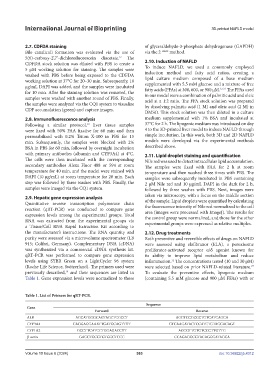
            previously described,  and their sequences are listed in   To evaluate the preventive effects, lipogenic medium
                             19
            Table 1. Gene expression levels were normalized to those   (containing 5.5 mM glucose and 900 μM FFAs) with or


            Table 1. List of Primers for qRT-PCR.
                                                               Sequence
             Gene
                                          Forward                                    Reverse
             ALB                 AGCATGGGCAGTAGCTCGCCT                      AGGTCCGCCCTGTCATCAGCA
             CYP3A4             CAGGAGGAAATTGATGCAGTTTT                  GTCAAGATACTCCATCTGTAGCACAGT
             CYP1A2               GCCTTCATCCTGGAGACCTT                       AGCGTTGTGTCCCTTGTTG
             β-actin               GAGCTGCGTGTGGCTCCC                      CCAGAGGCGTACAGGGATAGCA


            Volume 10 Issue 6 (2024)                       363                                doi: 10.36922/ijb.4312

