
Page 374 - IJB-10-6
P. 374
International Journal of Bioprinting 3D-printed NAFLD model
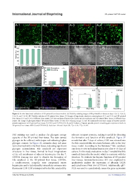
Figure 2. In vitro functional validation of 3D-printed liver tissue models. (A) Viability staining images of 3D-printed liver tissue on days 1 (A-1), 3 (A-2),
5 (A-3), and 7 (A-4). (B) Viability statistics of 3D-printed liver tissue. (C) Images of hepatocyte clusters in microspheres (C-1 and C-2) and 3D-printed
liver tissue (C-3 and C-4) at different time points. (D) Size analysis of hepatocyte clusters in microspheres and 3D-printed liver tissue at different time
points. (E) Image of glycogen-stained 3D-printed liver tissue. (F) Bile duct staining image. (G and H) Immunofluorescence image of hepatic-specific
protein expression in 3D-printed liver tissue: ALB (G) and CYP3A4 (H). (I and J) Analysis of hepatic-specific protein-encoding gene expression levels in
3D-printed liver tissue: ALB (I) and CYP3A4 (J). Scale bar: 100 and 10 µm (E). **p < 0.01; ***p < 0.001.
PAS staining was used to analyze the glycogen storage relevant transport proteins, making it useful for detecting
capacity of the 3D-printed liver tissue. The stain turned the formation and function of bile canaliculi. Figure 2F
glycogen in the cells red, with deeper red indicating higher reveals that after 7 days of culture, CDF was released into
glycogen content. In Figure 2E, extensive deep red areas the bile canaliculi-like structures between cells in the liver
were observed within the liver tissue, indicating significant tissue model. According to the literature, bile canaliculi
39
glycogen accumulation. Bile canaliculi are important can form in vitro in human hepatocytes after 6–10 days of
structures in liver tissue, formed by local invaginations culture. In this study, evaluation on day 7 revealed that bile
of the cell membranes of adjacent hepatocytes. On day 7, canaliculi formed in the model, which is consistent with the
CDFDA staining was used to observe the formation of literature. To evaluate the hepatic function of 3D-printed
bile canaliculi in the 3D-printed liver tissue. CDFDA, liver tissues, immunofluorescence (IF) was employed to
a nonfluorescent, nonpolar ester compound, enters qualitatively analyze the expression of albumin (ALB)
hepatocytes and is hydrolyzed by intracellular esterases into and cytochrome P450 3A4 (CYP3A4). Additionally, qRT-
fluorescent CDF. CDF is transported to the bile canaliculi by PCR was used to quantitatively assess the gene expression
Volume 10 Issue 6 (2024) 366 doi: 10.36922/ijb.4312