
Page 433 - IJB-10-6
P. 433
International Journal of Bioprinting 3D-bioprinted respiratory disease model
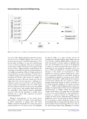
Figure 13. Viral growth curve of influenza A virus (IAV) determined by plaque assay for static, dynamic, and dynamic with nanoparticle culture conditions.
that some of the collagen and gelatin materials may have the bioink’s ability to co-culture primary cells for an
leached out of the crosslinked alginate material due to the extended time with high viability. Other studies reported
extended period spent at elevated temperatures (37°C) in the literature, utilizing slightly different materials and
during culture, resulting in the mass loss and downward A549 cells instead of primary respiratory epithelial cells,
trend in mechanical properties. This may be reduced by have also demonstrated that the use of 3D-bioprinted
crosslinking at a higher concentration for a greater length co-culture can extend cell culture life beyond 28 days. 41,42
of time to form a tighter polymeric mesh; however, this Along with the use of primary respiratory cells in the
would have to be balanced with the possible decrease in form of HPFs and HBEpCs for increased physiological
cell viability post-printing. While the Young’s modulus of relevance of the model, the incorporation of other
the printed lattice structure sat between 15 and 25 kPa, it methods for increasing relevance, including the culture
is within the range of that measured for native lung tissue of the bioprinted constructs in a bioreactor mimicking
throughout the 28-day culture period. In contrast, the the biomechanical stimulus of the lung increased cellular
compressive modulus of the bulk material and UTS are proliferation/metabolism. Additionally, the incorporation
higher than those of native lung tissue. Although these of HGF-loaded nanoparticles also resulted in a further
51
mechanical studies were carried out in cell-free scaffolds, (though insignificant) increase in cellular metabolism at
previous studies demonstrated that very similar values the 28-day timepoint, building on the results of previous
were found between cell-free and cell-containing scaffolds studies. 33,34 While live/dead imaging helped confirm the
over a 14-day period. This similarity likely results from presence of a high density of viable cells, the background
cell remodeling, which balances material degradation autofluorescence of collagen within the green “live”
caused by cells with the deposition of cellular matrix channel created noise in the images that was challenging
materials, but it cannot fully compensate for general to fully remove without significant image manipulation.
material degradation. 34 Therefore, an improved process for imaging may be
required for future studies. While live/dead imaging did
Biological characterization of the bioink further qualitatively confirm the increased cell density indicated
demonstrates its ability to be used in RTE applications, by the quantitative cellular metabolism studies, it also
as cellular metabolism of printed HPFs and seeded demonstrated that the incorporated cells did not take on
HBEpCs measured through the XTT assays was seen to physiologically relevant morphology. This may be due to
steadily increase over the 28-day period. This highlights the encapsulating bioink being challenging to remodel,
Volume 10 Issue 6 (2024) 425 doi: 10.36922/ijb.3895