
Page 454 - IJB-10-6
P. 454
International Journal of Bioprinting 3D-printed scaffold for biomolecule delivery
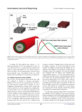
Figure 1. Experimental illustration of the 3D scaffold for the delivery of dual biomolecules. (a) The 3D printing and coating processes. (b) Expected release
of growth factors from the double layers. Abbreviations: PCL: Poly-ε-caprolactone; GS: Gelatin-silica; bFGF: Basic fibroblast growth factor; BMP-2: Bone
morphogenetic protein-2.
To release GFs, the scaffolds were soaked in 1 mL formed a monolayer. Passages three and four were used
PBS and incubated at 37°C with shaking for 42 days. At for further evaluation. To confirm the biological effects
a pre-determined time, the supernatant was collected and of bFGF and BMP-2 released from the bilayers on the
stored at −80°C until further use. The release profiles of scaffolds, initial adhesion and viability in conditioned and
bFGF and BMP-2 were assessed using an enzyme-linked osteogenic media were examined. The activated scaffolds
immunosorbent assay (Quantikine ELISA kit; R&D were sterilized with 70% ethanol and ultraviolet light.
5
Systems, USA) according to the manufacturer’s instructions. For the adhesion assay, 1 × 10 cells were seeded onto the
scaffolds and incubated for 2 h. The cell adhesion samples
2.3. Cellular responses on the scaffolds were fixed with 4% paraformaldehyde solution for 15
The rat bone marrow-derived mesenchymal stem cells min and permeabilized in 0.1% Triton X-100 in PBS for
were obtained as detailed in previous experiments. 10 min. The cytoskeletons and nuclei were stained with
Briefly, the bone marrow was separated from the femora phalloidin and Hoechst 33342, respectively, in PBS and
and tibia (age four weeks) with 2.5 mg/mL of type I were visualized using CLSM. Cell viability was measured
collagenase (ThermoFisher Scientific, USA). Single cells in stem cells cultured on scaffolds for 14 days using a
were harvested from the cell suspension by centrifugation cell counting kit-8 (CCK; Dojindo Laboratories, Japan)
and sieved three times. The cells were cultured in total according to the manufacturer’s instructions.
culture medium, which is α-Minimum Essential Medium To confirm the osteogenic ability of BMP-2 released from
(α-MEM; Welgene, Korea) supplemented with 10% fetal the scaffolds, cells were cultured for 24 days in osteogenic
bovine serum and 1% penicillin/streptomycin (Gibco, media consisting of conditioned media supplemented with
USA) in a humidified atmosphere of 95% air and 5% CO ascorbic acid (50 μg/mL) and β-glutaraldehyde (10 mM).
2
at 37°C. Nonadherent cells were removed by the medium For alkaline phosphatase (ALP) activity measurement,
exchange when the cells reached 70% confluence and cell lysates extracted from the samples cultured for seven
Volume 10 Issue 6 (2024) 446 doi: 10.36922/ijb.4638