
Page 457 - IJB-10-6
P. 457
International Journal of Bioprinting 3D-printed scaffold for biomolecule delivery
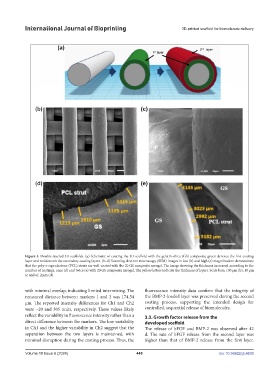
Figure 3. Double-layered 3D scaffolds. (a) Schematic of coating the 3D scaffold with the gelatin-silica (GS) composite; green denotes the first coating
layer and red denotes the secondary coating layers. (b–d) Scanning electron microscopy (SEM) images in low (b) and high (c) magnification demonstrate
that the poly-ε-caprolactone (PCL) struts are well-coated with the 20 GS composite xerogel. The image showing the thickness increased according to the
number of coatings, once (d) and twice (e) with 20 GS composite xerogel. The yellow letters indicate the thickness of layers. Scale bars: 100 µm (b); 10 µm
(c and e); 2 µm (d).
with minimal overlap, indicating limited intermixing. The fluorescence intensity data confirm that the integrity of
measured distance between markers 1 and 2 was 174.54 the BMP-2-loaded layer was preserved during the second
μm. The reported intensity differences for Ch1 and Ch2 coating process, supporting the intended design for
were −20 and 305 units, respectively. These values likely controlled, sequential release of biomolecules.
reflect the variability in fluorescence intensity rather than a 3.3. Growth factor release from the
direct difference between the markers. The low variability developed scaffold
in Ch1 and the higher variability in Ch2 suggest that the The release of bFGF and BMP-2 was observed after 42
separation between the two layers is maintained, with d. The rate of bFGF release from the second layer was
minimal disruption during the coating process. Thus, the higher than that of BMP-2 release from the first layer.
Volume 10 Issue 6 (2024) 449 doi: 10.36922/ijb.4638