
Page 458 - IJB-10-6
P. 458
International Journal of Bioprinting 3D-printed scaffold for biomolecule delivery
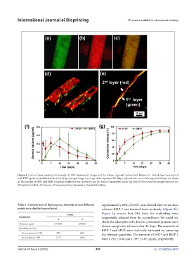
Figure 4. Confocal laser scanning microscope (CLSM) fluorescence images and the release of growth factors from bilayers. (a–e) Each layer was stained
with FITC (green; a) and rhodamine (red; b) dye; merged image (c); image of the separated 3D bilayer (d); and side-view of the separated bilayer (e). (f and
g) The amount of bFGF and BMP-2 released at different time points (f) and the ratio of cumulative release (g) from 20 GS composite xerogel layers on the
3D-printed scaffold. All data (n = 8) are presented as the mean ± standard deviation.
Table 1. Comparison of fluorescence intensity at two different Approximately 60% of bFGF was released after seven days,
points on a double-layered strut. whereas BMP-2 was released twice as slowly (Figure 4f).
Point Figure 4g reveals that GFs from the scaffolding were
Parameter sequentially released from the second layer. We could not
1 2
Distance (μm) 175.06 349.60 check the adsorptive GFs, but the pretreated proteins were
almost completely released after 42 days. The amounts of
Intensity (units)
BMP-2 and bFGF were indirectly estimated by summing
Green channel, Ch1 903 883 the released quantities. The amounts of bFGF and BMP-2
Red channel, Ch2 1151 1456 were 1.792 ± 0.86 and 1.700 ± 0.87 µg/mL, respectively.
Volume 10 Issue 6 (2024) 450 doi: 10.36922/ijb.4638