
Page 15 - IJB-7-4
P. 15
Zhuang, et al.
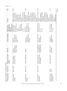
Ref [121] [120] [122] [123] (Contd...)
High cell viability in mono- and co-culture More resistant to paclitaxel than individual cells Size limitation of Limited control over spheroid arrangement MDA-MB-231 migrates out of spheroids in co-culture with HUVECs Limited control on bioprinted spheroid location and number Size limitation of Reduction of the lipid Increased fibronectin, collagen I and collagen Limited control over spheroid arrangement Size l
Feature • • • spheroid • • • • spheroid • content • VI expression • • spheroid • • spheroids
Spheroid size/ spheroidization time (ST)/ fusion time (FT) 5000 cells/well Size~100 µm ST: MCF10A cells, 8–10 d; MCF-7, MDA-MB-231, MCF10A-NeuN, 5–6 d FT: N.A. Size<70 µm ST: MCF10A, 8 d; MDA-MB-231, 5 d FT: N.A. 4800 spheroids/mL Size: 228 ± 22 μm ST: 2 d FT: N.A. Size: 300/500 μm ST: A few minutes FT: 70 h
Cell type (density) MCF10A, MCF10A-NeuN, MDA-MB-231, MCF7 MCF10A MDA-MB-231 (10000 cells/well) HUVECs ASCs (2500 cells/ spheroid) MDA-MB-231 Cardiac and endothelial cells, VEGF
Materials Matrigel, gelatin-alginate, collagen-alginate Matrigel Thiol-modified HA, unmodified high concentration HA, Collagen type I (Bio-paper)
Spheroid generation method Falcon 8 chamber polystyrene vessel 8-well chamber slide Agarose molds cast in MicroTissues®3D Petri Dishes® Pellet centrifugation
Table 1. 3D printing assisted spheroid assembly Target tissue Printing strategy Breast cancer Extrusion-based Breast cancer Extrusion-based Breast cancer Extrusion-based Cardiac Extrusion-based tissue printing (capillary
bioprinting
printing
printing International Journal of Bioprinting (2021)–Volume 7, Issue 4 micropipette) 11