
Page 16 - IJB-7-4
P. 16
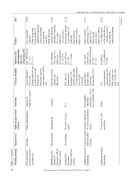
Using Spheroids to build 3D Bioprinted Tumor Microenvironment


               Ref       [138]                 [139]       [131]            [137]          [135]            (Contd...)


                         Size consistent   • Long fusion time  Large quantity of   spheroids preparation   is time consuming  Non-uniform   • Weak spheroids  Turnstile allows the   deposition of spheroid   one at a time  vascularization  Mechanical damage   to the integrity of   Fixed spacing   between needles  High resolution   positioning (~10%   spheroid size)  High density   micro-tissue  • High cell viability  ~11%with respect   to the spheroid size   --position accu


               Feature   •    spheroids  •    •    structure  •    Improved   •    •    spheroids  •    • High cost   •    •    •    •


               Spheroid size/  spheroidization   time (ST)/  fusion time (FT)  Size: 300/500 μm  ST: 1-2 h  FT: 5-7 d  Size: thyroid,    388.2 μm±45.3;  Allantoide,    493.6 μm±114.3  ST: 18-24 h  Size: 500 µm  ST: 48 h  FT: 3 weeks   Size: 5000   cells/200 µm  10000 cells/400 µm  ST: 96h  FT: 4d  80-800 µm  (~30 s/spheroid)  ST: 24h


               Cell type (density)  Chinese Hamster   Ovary cell,  Human umbilical vein   smooth muscle cells,  Human skin   fibroblasts, porcine   aortic smooth muscle   cells   Individual thyroid  explants and   allantoides  iPSC-derived   human neural   progenitor cells   (40,000 cells/well),  U118 human glioma   cells (10,000    cells/well)  Human MSCs,  Human cardiac   fibroblasts  Human iPSC-CM  3T3,  mouse mammary   carcinoma line 4T1,  HUVECs/MSCs,  HDF, electrocy


               Materials  Agarose as   temporary support  Collagen  N.A.    HA modified   with either   adamantane (Ad) or   β-cyclodextrin (CD)  Fibrin


               Spheroid generation   method  Pellet centrifugation  Hanging drop  96-well U-bottom   plates  Ultra-low attachment   96-well round-bottom   plates  U-bottom 96-well   microplate


               Target tissue  Vascular         Thyroid gland  Glioblastoma  Post-myocardial   infarction (MI)   scarring  /


            Table 1. (Continued)  Printing strategy  Extrusion-based   printing (capillary   micropipette)  Multifunctional   Fabion 3D   bioprinter with the   turnstile system  Scaffold-free   bioprinter/  Regenova/kenzan   method  Aspiration-assisted   bioprinting  Aspiration-assisted   bioprinting


           12                          International Journal of Bioprinting (2021)–Volume 7, Issue 4