
Page 76 - IJB-7-4
P. 76
Bioink for Reconstruction of Rigid-living Systems
The rise in the orange box, at 50 ppm, and the green integration as the methacrylate ions can be observed
box, between 35 and 25 ppm, disappears when there between 6 and 6.5 ppm [28,29].
are bioceramics; therefore, these signals are a piece of In the TGA (Figure 5A), there is a reduction
evidence from the interaction of binding from inorganic of 13% of weight from 90°C to 100°C due to the loss
components of calcium, phosphate, and carbonate ions to of H O-coordinated ions remaining in the crystalline
2
the biopolymer side from the formulation. This data can arrangements of ceramics compressed with the polymer.
be corroborated in future studies with P MAS NMR and From 100°C to 642°C, there is a loss of 10% from the
31
43 Ca MAS NMR [27,28] . Besides, in H-NMR (Figure S6), it sample, equivalent to the biopolymers that were calcined
1
can be complemented the presence of the methacrylation under this procedure; this variation comes from the
functionalization of the GelMA synthesis and PEGDA different polymeric ionic/photo-crosslinking behaviors
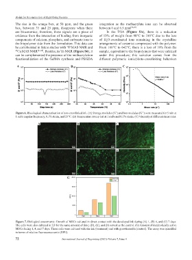
A B C
Figure 6. Rheological characterization of non-crosslinked ink. (A) Storage modulus (G’) and loss modulus (G’’) were measured for 5 min at
1 rad/s angular frequency, 0.1% strain, and 25°C. (B) Temperature sweep test at 1 rad/s and 0.1% strain. (C) Viscosity at different shear rates.
A B C
D E F
G
Figure 7. Biological assessments. Growth of MSCs cultured in direct contact with the developed ink during (A) 1, (B) 4, and (C) 7 days.
The cells were also cultured in 2D for the same amount of time; (D), (E), and (F) served as the control. (G) Amount of metabolically active
MSCs during 1, 4, and 7 days. These cells were cultured with the ink (treatment) and with growth media (control). The assay was quantified
in terms of relative fluorescence units (RFU).
72 International Journal of Bioprinting (2021)–Volume 7, Issue 4