
Page 8 - IJB-7-4
P. 8
Using Spheroids to build 3D Bioprinted Tumor Microenvironment
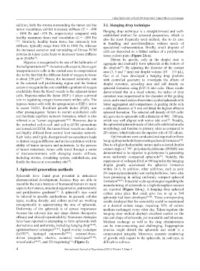
addition, both the stroma surrounding the tumor and the 3.1. Hanging drop technique
tumor vasculature exhibit increased stiffness (E = ~800 Hanging drop technique is a straightforward and well-
– 1000 Pa and ~450 Pa, respectively) compared with established method for spheroid preparation, which is
healthy mammary tissue and vasculature (E = ~200 Pa)
[32] . Similarly, healthy brain tissue has extremely low also the most frequently used method, due to its ease
in handling and user-friendliness without needs of
stiffness, typically range from 100 to 1000 Pa, whereas specialized instrumentation. Briefly, small droplets of
the increased secretion and remodeling of fibrous ECM cells are deposited on a lidded surface of a polystyrene
proteins in tumor niche leads to increased tissue stiffness tissue culture plate (Figure 2A-a).
up to 26 kPa . Driven by gravity, cells in the droplet start to
[33]
Hypoxia is recognized to be one of the hallmarks of aggregate and eventually form spheroids at the bottom of
the malignant tumors . As tumor cells expand, the oxygen the droplets . By adjusting the diameter of the contact
[34]
[52]
transportation to cells in the central zone is compromised area (3, 5 and 7 mm) and droplet volume (10 – 153 µL),
due to the fact that the diffusion limit of oxygen in tissue Gao et al. have developed a hanging drop platform
is about 250 µm . Hence, the increased metabolic rate with controlled geometry to investigate the effects of
[35]
in the external cell proliferating region and the limited droplet curvature, spreading area and cell density on
access to oxygen in the core establish a gradient of oxygen spheroid formation using β-TC-6 islet cells. These results
availability from the blood vessels to the adjacent tumor demonstrated that at a fixed volume, the radius of drop
cells. Hypoxia‐inducible factor (HIF-) 1 plays a pivotal curvature was proportional to the diameter of the guiding
role in regulating oxygen homeostasis within cells. The circle, and a small radius of curvature yielded spheroids with
hypoxic tumor cells with the upregulation of HIF-1 strive better aggregation and compactness. A guiding circle with
to secret VEGF, fibroblast growth factor (FGF), and a selected diameter of 5 mm exhibited the highest efficient
other proangiogenic factors to recruit endothelial cells spheroid formation. The selected cell density of 105 cells/
and facilitate capillary network formation, which is also mL gave rise to spheroids with a diameter of 400 – 500 µm,
referred to as “tumor angiogenesis” . However, due to which was well aligned with native islet size . Notably,
[53]
[36]
the perturbed cell-to-cell and cell-to-ECM interactions the optimized spheroids made of islet cells exhibited similar
and remodeled ECM, the tumor blood vessels are chaotic morphology and function to primary islets as compared to
and highly differed from normal host vascular network. 2D culture, which indicates the superior role of 3D culture.
Such leaky and highly disorganized neovasculature leads Optimizations were conducted and reported on these
to limited oxygen diffusion and are in correlation with the hanging drop methods for promoting spheroid formation .
[38]
ability of tumor invasion and metastasis. In the process Due to a higher hydrophobic nature and a selected droplet
of tumor metastasis, tumor cells travel through a series contact angle of 99°, polydimethylsiloxane (PDMS) was
of microenvironment with changing matrix stiffness, demonstrated to be superior to polystyrene in generating
[52]
including stroma, circulating system, endothelium, and more uniformly compacted spheroids . Notably, the
finally the tissues at a secondary site . supplement of collagen fibril at 500 µg/ml in the hanging
[37]
droplet greatly accelerated the spheroid formation
3. Spheroid generation methods within 24 h. In addition, other additives, such as poly
(N-isopropylacrylamide) and methylcellulose, have also
Spheroids have found great potential in anticancer been promising in aiding uniformly compact spheroid
pharmaceutical development, because they are able to formation [54,55] . Industrial scale-up strategies regarding the
resemble the main features of humanoid tumors in many manufacturing of spheroids in a high-throughput manner
aspects; for instance, structural organization, and metabolic are reported (Figure 2A-a ). A hanging drop spheroid
and proliferative gradients . A spheroid’s size could culture array plate that could give rise to up to 384
1
[13]
be tailored to specific applications. In general, cellular spheroids had been developed [39,40] . These corresponding
types, seeding density, and culture period are working results displayed that the osmolality could be maintained
synergistically in appropriating the size of spheroids. at a desired culture range, requiring 30% of culture
Uniformity of the spheroids is of utmost importance medium exchanged every other day. Taken together, the
because the relevant size and shape dictate therapeutic hanging drop method displays excellent control on the
efficacy and clinical reproducibility. Numerous strategies size and shape of spheroids, yet is unstable and laborious.
have been reported in attempting to form spheroids with Medium exchange as well as the drug administration
desired size and uniformity, including hanging drop [38-40] , can be time-consuming and challenging. Inappropriate
agitation‐based techniques [41,42] , liquid overlay technique practice might disturb the spheroids and result in a
(LOT) , hydrogel microwells [44,45] , external-force- compromised integrity. Moreover, accurate monitoring
[43]
driven (magnetic, electric, acoustic) techniques [46-48] , of growth with regard to the spheroids, in real-time, is
microfluidics [49,50] , and 3D bioprinting (Figure 2). difficult to achieve.
[51]
4 International Journal of Bioprinting (2021)–Volume 7, Issue 4