
Page 84 - IJB-8-1
P. 84
Geometric Accuracy of 3D Printed Dental Implant
A
B
C
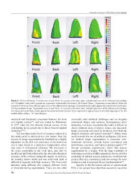
Figure 5. (A) Overall Group - Printed versus Actual Tooth. An example of the front, back, left and right views of the differences in topology
of a 3D-printed tooth (solid) against the segmented tooth model (wireframe). (B) Printed Group - Segmented versus Printed Tooth. An
example of the front, back, left and right views of the differences in topology of a printed tooth (solid) against the actual tooth (wireframe).
(C) Segmentation Group - Segmented versus Actual Tooth. An example of the front, back, left and right views of the differences in topology
of a segmented tooth model (solid) against the actual tooth (wireframe). Green represents a high accuracy at which the topology of the two
models differs within a +0.1 mm tolerance.
structural and functional connection between the bone commonly cited technical challenges such as irregular
and implant surface , and was coined by Brånemark anatomical shapes and surfaces, heterogeneous pixel
[23]
in 1977 when he first showed clinical success of the intensities, and noisy boundaries that make it difficult to
oral implant in his patient due to direct bone-to-implant clearly delineate areas of interest. Hence, any inaccurate
anchorage [24,25] . image processing will result in deviation error from the
The less-than-desired level of accuracy achieved in planned treatment and desired outcome . Future work
[26]
this study could be attributed to a few factors. First, the could consider the use of artificial intelligence (AI) tools
success of image segmentation by thresholding is highly for medical image segmentation to overcome inter-
dependent on the skill and experience of the technician technician variations, provide higher consistency and
and is often based on a subjective interpretation which performance outcomes, and improve product quality [27,28]
may result in inter-person variations. The resolution of through automated segmentation, rather than manual
the scans, particularly at the tooth apex, may also be segmentation by a human. With the large variability of
insufficient and further limited by the allowable threshold tooth shapes and sizes of different patients, the use of AI for
tolerance of the software during file import. In addition, prediction of optimal process parameters would provide
the monkey incisor tooth root was small and made it greater efficiency, consistency, and cost-savings for mass
difficult to segment with high accuracy. The bone-tooth production and widespread clinical implementation .
[10]
densities being different also required different levels Specific to the fabrication and use of custom-made
of threshold during segmentation. There are also other RAI, a few groups have described successful outcomes
70 International Journal of Bioprinting (2022)–Volume 8, Issue 1