
Page 26 - IJB-8-2
P. 26
Bioprinting Gelatin-Recombinant Type III Collagen Hydrogel for Wound Healing
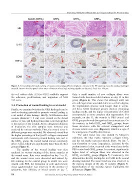
Figure 3. Immunohistochemical staining of tissues surrounding different implants. Arrows in the HE staining show the residual hydrogel
material. Arrows in other panels show areas of interest where high staining signals are detected. Scale bar: 100 μm.
the cell culture dish. All five GRH scaffolds support Only a small number of new collagen fibers were
the adhesion, proliferation, and migration of NIH formed with disordered distributions on day 13 in this
3T3 cells. group (Figure 6). This shows that although adult rats
can self-regenerate wounded skins to a certain degree,
3.4. Promotion of wound healing in a rat model the regeneration process took longer than 2 weeks.
Finally, we examined whether the GRH hydrogels can be All three GRH treatment groups showed promising
used as dressing materials to promote wound healing in healing results, and the higher concentration of rColIII
a rat model of skin damage. Briefly, full-thickness skin corresponded to more complete skin regeneration. For
wounds (diameter = 2 cm) were created on the dorsal example, on day 13, the wounds in PBS control and
surface of rats, and hydrogel materials were then applied GRH groups showed disorganized tissue structures. On
0
to the surface of the wounds. After a designated duration the contrary, in both GRH and GRH groups, dense
10
20
of incubation, the skin samples were removed and tissue structures with the ordered arrangement and
analyzed by various methods. First, the wound sizes in obvious cuticle were seen (Figure 6), which is a sign of
different groups were recorded. We observed a trend that the emergence of healthy skin tissues.
the higher percentage of the type III collagen component The same tissue was also stained by Masson’s
corresponds with increasing wound healing rate, and in trichrome; the blue marks represent collagen fibers,
the GRH group, the wound healed almost completely and the red marks represent muscle fibers. On day 7,
20
after 13 days, which was significantly faster than all other scar formation or tissue hyperplasia, indicated by the
groups (Figure 5). predominant red color, occurred at the wound area in the
The quality of the wound healing was then PBS control and GRH groups. In contrast, significantly
0
analyzed by various staining of the tissue samples at higher level of collagen fibers was observed in the
the wound areas. Rats were sacrificed on day 7 or 13, GRH and GRH groups (Figure 7). Consistently, this
10
20
and the wound areas were dissected and stained by demonstrates healthy healing due to rColIII in the wound
HE staining. In the control group in which rats were dressing material. The same trend was also observed in
treated with PBS, the formation of scab and necrotic the staining images of the day 13 samples (Figure 7).
tissue agglomeration was observed in the absence of Quantification of the staining signals highlighted the
epidermis, hair follicles, or blood vessels on day 7. differences (Figure 7B).
18 International Journal of Bioprinting (2022)–Volume 8, Issue 2