
Page 25 - IJB-8-2
P. 25
Huang, et al.
A B
C
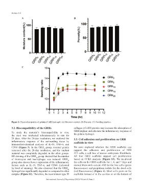
Figure 2. Physical properties of gelatin rColIII hydrogel. (A) Moisture content. (B) Porosity. (C) Swelling kinetics.
3.2. Biocompatibility of the GRHs collagen (rColIII) protein may increase the absorption of
GRH implant and alleviate the inflammatory response of
To study the material’s biocompatibility in vivo, the gelatin hydrogel.
the stent was implanted subcutaneously in rats for
28 days. After the 28-day incubation, we explored the 3.3. Cell adhesion and proliferation on GRH
inflammatory response of the surrounding tissue by scaffolds in vitro
immunohistochemical analyses of IL-10, TNF-α, and
CD68 (Figure 3). In the GRH group, residual gelatin We next explored whether the GRH scaffolds can
0
remained after the 28-day incubation, and the implant support the adhesion and proliferation of NIH
material was completely degraded in the other groups. 3T3 cells, a cell line of mouse embryonic fibroblasts.
HE staining of the GRH group showed that the number All four GRH scaffolds support cell proliferation
20
of monocytes and macrophages was reduced. GRH based on CCK8 analysis (Figure S3). We incubated
20
group also showed lower expression of the inflammatory the cells on the GRH scaffolds for 1, 4, and 7 days and
factors such as IL-10, TNF-α, and CD68 (indicated stained them with calcein AM for the live cells (green
by level of staining). We also observed that the GRH fluorescence) and propidium iodide for the dead cells
20
hydrogel was significantly degraded as compared to other (red fluorescence) (Figure 4). Most cells grew on the
samples (Figure S2). Therefore, the recombinant type III scaffolds instead of in the cavities or on the bottom of
International Journal of Bioprinting (2022)–Volume 8, Issue 2 17