
Page 90 - IJB-8-2
P. 90
3D-printed Stent Coated with Dipyridamole-loaded Nanofiber
A B
C D
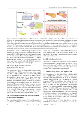
Figure 1. Fabrication of a 3D-printed bioresorbable stent coated with dipyridamole (DP)-loaded nanofiber and its mechanism for restenosis
prevention and endothelialization. (A) Fabrication and implantation of stents coated with DP-loaded dipyridamole-loaded poly(D,L-lactide)
(PDLLA) nanofibers, (i) 3D-printed stents were prepared using a 3D printing system with a rotation mandrel, (ii) PDLLA nanofibers
loaded with DP were randomly deposited onto 3D-printed stents, and (iii) stents were implanted into porcine coronary arteries for in vivo
evaluation. (B) The artery segment implanted with the bare stent exhibited more severe in-stent restenosis, while the artery implanted with
DP-loaded stents showed initial endothelialization. (C) The proposed mechanism of stents coated with DP-loaded nanofibers for antiplatelet
adhesion, the inhibition of smooth muscle cells proliferation, and enhanced endothelial cell growth.
3D-printed PCL stents. A similar electrospinning process was of DP, PDLLA nanofibers, and PDLLA/DP nanofibers
introduced in our previous work . With the rotation of the was examined by FTIR spectroscopy (Bruker V70,
[23]
mandrel, PDLLA/DP nanofibers were uniformly deposited Germany). The scanning wavenumber range was from
onto 3D-printed stents. Thus, 3D-printed PCL stents coated 4000 cm to 600 cm .
−1
−1
with PDLLA/DP nanofibers were accomplished. To further
demonstrate the function of PDLLA/DP nanofibers, bare 2.5. Electrical conductivity
stents and stents coated with only PDLLA nanofibers The electrical conductivity of different polymer solutions
(without DP) were prepared as control groups. was measured using a conductometer (DDS-11A, INESA
2.3. Morphological and mechanical Scientific Instrument Co., Ltd., Shanghai, China). 5 mL
characterization of stents of each polymer solution was measured at 25°C. Three
samples (n = 3) from each group were used.
The surface morphology of 3D-printed PCL stents, stents
coated with only PDLLA nanofibers, and stents coated 2.6. In vitro drug release and degradation
with PDLLA/DP nanofibers was characterized by SEM To investigate the in vitro drug release behavior of DP
(ZEISS GeminiSEM 300, Germany). To further observe from PDLLA/DP nanofibers, weighted electrospun
the morphology of the inside view of stents, stents were nanofiber mats were immersed in 10 ml of phosphate-
cut in half from the middle before SEM characterization. In buffered saline (PBS, pH 7.4). The samples (n = 6) were
this study, the crush resistance test using parallel plates was incubated at 37°C. At predefined time points, 10 mL of
conducted by a mechanical test instrument (BOSE 3230, PBS solution with the released drug was collected, and
Germany). The stents used in this test possessed an inner 10 mL of fresh PBS was replenished. The absorbance
diameter of 3 mm and a length of 10 mm. The stent was value of PBS with released DP was recorded at 284 nm
placed between two plates and compressed to a displacement by an ultraviolet-visible spectrophotometer (HITACHI
of 1.5 mm (half of the stent inner diameter) at a constant U3900, Japan). To plot the standard curve of DP
moving speed of 1 mm/min. For each group, three samples concentration versus absorbance in PBS, the absorbance
were used to record the “Force-Displacement” curve. of different solutions with concentrations from 1 μg/mL
2.4. Morphological and FTIR characterization to 100 μg/mL was measured. To obtain the whole drug
of electrospun nanofibers release result, the absorbance value was accumulated
based on the previous values.
The morphology of PDLLA and PDLLA/DP nanofibers To determine the in vitro degradation behavior,
was obtained by SEM observation. The chemical bonding PDLLA/DP nanofibers were immersed in PBS, and
82 International Journal of Bioprinting (2022)–Volume 8, Issue 2