
Page 94 - IJB-8-2
P. 94
3D-printed Stent Coated with Dipyridamole-loaded Nanofiber
A B
C D E
F
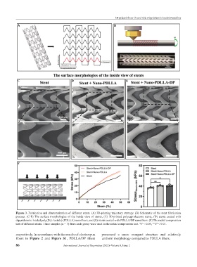
Figure 3. Fabrication and characterization of different stents. (A) 3D-printing trajectory strategy. (B) Schematic of the stent fabrication
process. (C-E) The surface morphologies of the inside view of stents, (C) 3D-printed polycaprolactone stents, (D) stents coated with
dipyridamole-loaded poly(D,L-lactide) (PDLLA) nanofibers, and (E) stents coated with PDLLA/DP nanofibers. (F) The radial compression
test of different stents. Three samples (n = 3) from each group were used in the radial compression test. *P < 0.05, **P < 0.01.
respectively. In accordance with the results of electrospun possessed a more compact structure and relatively
fibers in Figure 2 and Figure S1, PDLLA/DP fibers uniform morphology compared to PDLLA fibers.
86 International Journal of Bioprinting (2022)–Volume 8, Issue 2